. 2017 May;13(5):3889-3895.
doi: 10.3892/ol.2017.5896. Epub 2017 Mar 23.

Fibronectin induces epithelial-mesenchymal transition in human breast cancer MCF-7 cells via activation of calpain

Affiliations

Fibronectin induces epithelial-mesenchymal transition in human breast cancer MCF-7 cells via activation of calpain

Cheng-Lin Li et al. Oncol Lett. 2017 May.

Abstract

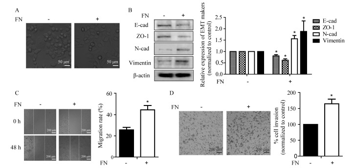
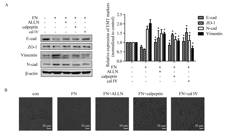
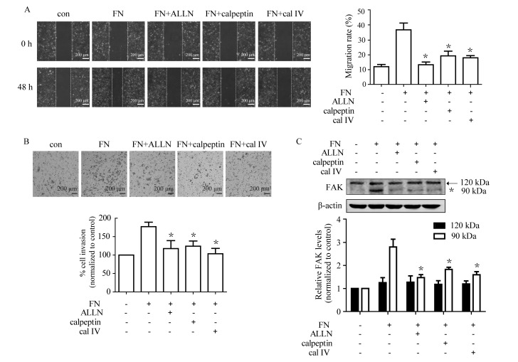
Fibronectin (FN) is a primary component of the mammary mesenchymal compartment and undergoes dramatic changes during breast cancer development. Increased FN expression is associated with an invasive and metastatic breast cancer phenotype. The present study demonstrated that FN causes an epithelial-mesenchymal transition (EMT)-like morphological change in MCF-7 breast cancer cells. FN stimulation caused the downregulation of epithelial markers E-cadherin and tight junction protein ZO-1, and the upregulation of mesenchymal markers N-cadherin and vimentin. Additionally, FN promoted cell migration and invasion in MCF-7 cells, with increased expression of calpain-2 and proteolysis of focal adhesion kinase 1 (FAK), indicating calpain activation. Notably, the FN induced changes in morphology and EMT markers were reversed with the treatment of calpain-specific inhibitors, calpain inhibitor I (N-acetyl-L-leucyl-L-leucyl-L-norleucinal), calpeptin and calpain inhibitor IV. Meanwhile, the effects of FN on cell migration and invasion, as well as FAK proteolysis were markedly suppressed by calpain inhibitors. Taken together, the results of the present study indicate that calpain plays an essential role in FN-induced EMT response, and that targeting calpain signaling may be a potential strategy to reduce breast cancer metastasis.

Keywords: breast cancer; calpain; epithelial-mesenchymal transition; fibronectin; invasion; migration.


Figures

Figure 1.
Figure 1.
FN stimulates changes in cell morphology and expression of EMT markers, as well as promoting cell migration and invasion in MCF-7 cells. (A) Micrographs of MCF-7 cells untreated and treated with FN for 48 h (magnification, ×400). (B) FN stimulated changes in the expression of EMT markers. (C) FN promoted migration of MCF-7 cells. Monolayer cells were scratched in the presence and absence of FN (20 µg/ml) for 48 h. The images were captured at 0 and 48 h following scratching (magnification, ×100). (D) FN promoted invasion of MCF-7 cells. Cells were plated with and without FN (20 µg/ml) for 48 h. The invasive cells that passed through the membrane were examined with crystal violet staining. The images are representative of 3 separate experiments (magnification, ×100). Values are expressed as the mean ± standard error. *P<0.05 vs. the control. E-cad, E-cadherin; FN, fibronectin; EMT, epithelial-mesenchymal transition; N-cad, N-cadherin; ZO-1, tight junction protein ZO-1.
Figure 2.
Figure 2.
Impact of FN on the processing of FAK and calpain expression in MCF-7 cells. (A) FN enhanced FAK proteolysis in MCF-7 cells. Arrow indicates the wild-type (120 kDa) and *indicates the processed form(s) (90 kDa) of FAK. (B) FN induced the upregulation of calpain-2 in MCF-7 cells. Values are expressed as the mean ± standard error. *P<0.05 vs. the control. FAK, focal adhesion kinase 1; FN, fibronectin.
Figure 3.
Figure 3.
Calpain mediates FN-induced EMT of MCF-7 cells. Cells were pretreated with and without ALLN (10 µM), calpeptin (50 µM) and calpain inhibitor IV (25 µM) for 1 h prior to treatment with FN for 48 h. (A) Inhibition of calpain activation blocked FN-induced change in EMT marker expression. The expression of EMT markers was detected by western blotting, with β-actin as loading control. Values are expressed the mean ± standard error. *P<0.05, FN vs. FN+ALLN, FN+calpeptin and FN+calpain inhibitor IV. (B) Repression of calpain activation inhibited FN-induced morphological changes. Morphological observation of MCF-7 cells was performed following FN treatment with or without calpain inhibitors (magnification, ×400). ALLN, calpain inhibitor I; con, control; EMT, epithelial-mesenchymal transition; FN, fibronectin; cal IV, calpain IV; E-cad, E-cadherin; N-cad, N-cadherin; ZO-1, tight junction protein ZO-1.
Figure 4.
Figure 4.
Calpain is involved in cell migration, invasion and FAK cleavage during FN-induced EMT in MCF-7 cells. Cells were pretreated with and without ALLN (10 µM), calpeptin (50 µM) and calpain inhibitor IV (cal IV, 25 µM) for 1 h prior to treatment with FN for 48 h. Cell migration and invasion, as well as processing of FAK, were subsequently analyzed. (A) The FN-induced cell migration was markedly suppressed by calpain inhibitors. (magnification, ×100) (B) FN-induced cell invasion was significantly inhibited by ALLN, calpeptin and calpain inhibitor IV. (magnification, ×100) (C) Pretreatment with calpain inhibitors suppressed FN-induced FAK processing. Values are expressed as the mean ± standard error. *P<0.05, FN vs. FN+ALLN, FN+calpeptin and FN+calpain inhibitor IV. ALLN, calpain inhibitor I; cal IV, calpain IV; EMT, epithelial-mesenchymal transition; FAK, focal adhesion kinase 1; FN, fibronectin; con, control.

References

    1. Oskarsson T. Extracellular matrix components in breast cancer progression and metastasis. Breast. 2013;22:S66–S72. doi: 10.1016/j.breast.2013.07.012. (Suppl 2) - DOI - PubMed
    1. Lu P, Weaver VM, Werb Z. The extracellular matrix: A dynamic niche in cancer progression. J Cell Biol. 2012;196:395–406. doi: 10.1083/jcb.201102147. - DOI - PMC - PubMed
    1. Ioachim E, Charchanti A, Briasoulis E, Karavasilis V, Tsanou H, Arvanitis DL, Agnantis NJ, Pavlidis N. Immunohistochemical expression of extracellular matrix components tenascin, fibronectin, collagen type IV and laminin in breast cancer: Their prognostic value and role in tumour invasion and progression. Eur J Cancer. 2002;38:2362–2370. doi: 10.1016/S0959-8049(02)00210-1. - DOI - PubMed
    1. Bae YK, Kim A, Kim MK, Choi JE, Kang SH, Lee SJ. Fibronectin expression in carcinoma cells correlates with tumor aggressiveness and poor clinical outcome in patients with invasive breast cancer. Hum Pathol. 2013;44:2028–2037. doi: 10.1016/j.humpath.2013.03.006. - DOI - PubMed
    1. Zhou Z, Qutaish M, Han Z, Schur RM, Liu Y, Wilson DL, Lu ZR. MRI detection of breast cancer micrometastases with a fibronectin-targeting contrast agent. Nat Commun. 2015;6:7984. doi: 10.1038/ncomms8984. - DOI - PMC - PubMed