

Identification of proteins with different abundance associated with cell migration and proliferation in leiomyoma interstitial fluid by proteomics
- PMID: 28521489
- PMCID: PMC5431234
- DOI: 10.3892/ol.2017.5943
Identification of proteins with different abundance associated with cell migration and proliferation in leiomyoma interstitial fluid by proteomics
Abstract
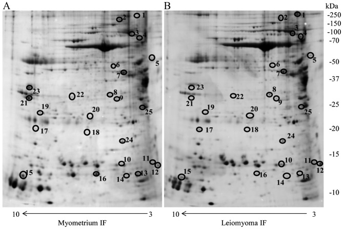
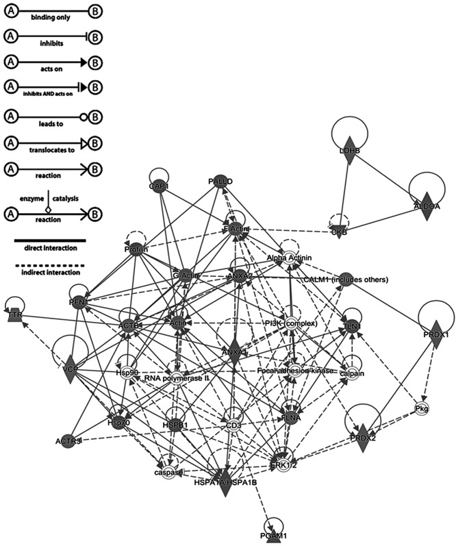
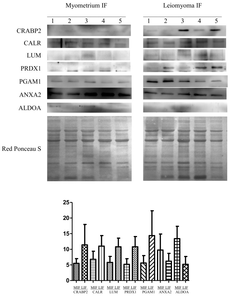
Uterine leiomyoma is the most common female reproductive tract benign tumor. Little is known about protein composition and changes in the leiomyoma interstitial fluid (IF). The present study focused on changes in protein abundance in the IF of leiomyoma. Leiomyoma IFs and adjacent myometrial IFs were obtained and analyzed by two-dimensional electrophoresis (2-DE) coupled with mass spectrometry and western blotting for 2-DE data validation. A total of 25 unique proteins were observed to change significantly (P<0.05). Of these proteins with different abundance, 22 had not been previously identified in leiomyoma IF. In silico analysis predicted that three of these proteins were secreted via classical mechanisms, while 22 were secreted via non-classical mechanisms. Ingenuity Pathway Analysis identified 17 proteins associated with cellular migration and proliferation. Among these, phosphoglycerate mutase 1 had not been previously associated with leiomyoma. The abundance of seven proteins was further validated by western blotting. A comparative proteomic approach identified a number of proteins associated with cellular migration and proliferation, with changes in abundance in IF likely to be involved in tumor development. Further studies will be required to investigate the role of these proteins in leiomyoma IF and their possible association with tumor development and growth.
Keywords: 2-D electrophoresis; interstitial fluid; leiomyoma; mass spectrometry; myometrium; proteomics; secreted protein.
Figures
Similar articles
-
Dysregulated chaperones associated with cell proliferation and negative apoptosis regulation in the uterine leiomyoma.Oncol Lett. 2018 May;15(5):8005-8010. doi: 10.3892/ol.2018.8325. Epub 2018 Mar 22. Oncol Lett. 2018. PMID: 29731911 Free PMC article.
-
A Proteomic Approach for the Identification of Up-Regulated Proteins Involved in the Metabolic Process of the Leiomyoma.Int J Mol Sci. 2016 Apr 9;17(4):540. doi: 10.3390/ijms17040540. Int J Mol Sci. 2016. PMID: 27070597 Free PMC article.
-
Two-dimensional gel electrophoresis analysis of the leiomyoma interstitial fluid reveals altered protein expression with a possible involvement in pathogenesis.Oncol Rep. 2015 May;33(5):2219-26. doi: 10.3892/or.2015.3827. Epub 2015 Mar 3. Oncol Rep. 2015. PMID: 25738828
-
Epidemiological and genetic clues for molecular mechanisms involved in uterine leiomyoma development and growth.Hum Reprod Update. 2015 Sep-Oct;21(5):593-615. doi: 10.1093/humupd/dmv030. Epub 2015 Jul 3. Hum Reprod Update. 2015. PMID: 26141720 Free PMC article. Review.
-
Tissue-specific stem cells in the myometrium and tumor-initiating cells in leiomyoma.Biol Reprod. 2014 Dec;91(6):149. doi: 10.1095/biolreprod.114.123794. Epub 2014 Nov 5. Biol Reprod. 2014. PMID: 25376230 Free PMC article. Review.
Cited by
-
The Deep Proteomics Approach Identified Extracellular Vesicular Proteins Correlated to Extracellular Matrix in Type One and Two Endometrial Cancer.Int J Mol Sci. 2024 Apr 24;25(9):4650. doi: 10.3390/ijms25094650. Int J Mol Sci. 2024. PMID: 38731868 Free PMC article.
-
Gel-Based Proteomic Identification of Suprabasin as a Potential New Candidate Biomarker in Endometrial Cancer.Int J Mol Sci. 2022 Feb 14;23(4):2076. doi: 10.3390/ijms23042076. Int J Mol Sci. 2022. PMID: 35216190 Free PMC article.
-
Phospho-DIGE Identified Phosphoproteins Involved in Pathways Related to Tumour Growth in Endometrial Cancer.Int J Mol Sci. 2023 Jul 26;24(15):11987. doi: 10.3390/ijms241511987. Int J Mol Sci. 2023. PMID: 37569364 Free PMC article.
-
Two Dimensional-Difference in Gel Electrophoresis (2D-DIGE) Proteomic Approach for the Identification of Biomarkers in Endometrial Cancer Serum.Cancers (Basel). 2021 Jul 20;13(14):3639. doi: 10.3390/cancers13143639. Cancers (Basel). 2021. PMID: 34298850 Free PMC article.
-
Dysregulated chaperones associated with cell proliferation and negative apoptosis regulation in the uterine leiomyoma.Oncol Lett. 2018 May;15(5):8005-8010. doi: 10.3892/ol.2018.8325. Epub 2018 Mar 22. Oncol Lett. 2018. PMID: 29731911 Free PMC article.
References
-
- Hashimoto K, Azuma C, Kamiura S, Kimura T, Nobunaga T, Kanai T, Sawada M, Noguchi S, Saji F. Clonal determination of uterine leiomyomas by analyzing differential inactivation of the X-chromosome-linked phosphoglycerokinase gene. Gynecol Obstet Invest. 1995;40:204–208. doi: 10.1159/000292336. - DOI - PubMed
LinkOut - more resources
Full Text Sources
Other Literature Sources
Miscellaneous