. 2017 May 18;19(1):100.
doi: 10.1186/s13075-017-1310-4.

M2 macrophage is the predominant phenotype in airways inflammatory lesions in patients with granulomatosis with polyangiitis

Affiliations

M2 macrophage is the predominant phenotype in airways inflammatory lesions in patients with granulomatosis with polyangiitis

Alexandre Wagner Silva de Souza et al. Arthritis Res Ther. .

Abstract

Background: Macrophages may present two distinct phenotypes indicated as M1 and M2 under different stimuli. M1 and M2 macrophages have divergent functions that range from enhancement of inflammation for M1 to tissue repair and remodeling for M2 macrophages. The objective of this study was to evaluate the distribution of M1 and M2 macrophage phenotypes in biopsies from the airways of patients with active granulomatosis with polyangiitis (GPA) and to analyze their associations with T and B cells in those biopsies, and with nasal carriage of Staphylococcus aureus, disease parameters and therapy.

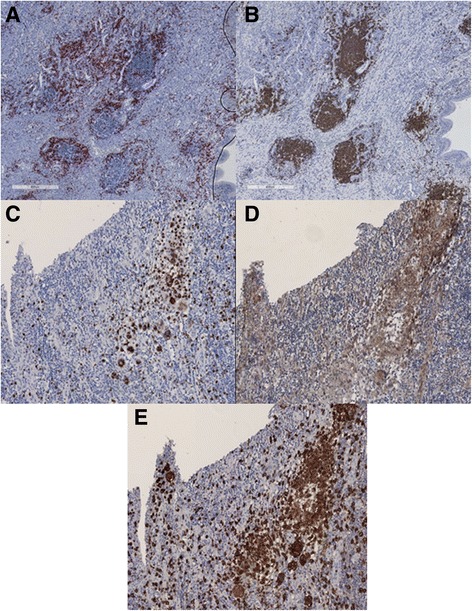
Methods: Consecutive GPA patients (n = 35) with active airway disease, who underwent respiratory tract biopsy were included. Immunohistochemical evaluation was performed to assess the distribution of macrophages and T and B cells using the markers CD68, CD3 and CD20, respectively. CD86 was used as the M1 marker and CD163 as the M2 marker while Tbet and GATA-3 were used as Th1 and Th2 markers, respectively. At the time of the biopsy patients were assessed for nasal carriage of Staphylococcus aureus and treatment.

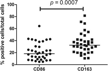
Results: Percentages of macrophages and T cells were significantly higher than those of B cells in lesional tissue from the respiratory tract in GPA. M2 macrophages and Th2 cells were more frequent than M1 macrophages (p = 0.0007) and Th1 cells (p < 0.0001), respectively. Percentages of T cells were higher in nose biopsies than in biopsies from other sites (p = 0.021); macrophages and CD163+ macrophages were more predominant in biopsy sites other than the nose (p = 0.039 and p = 0.012, respectively). Carriage of Staphylococcus aureus was associated with higher T cell scores (p = 0.014). The frequency of macrophages, especially M2 macrophages, was higher in GPA patients treated with immunosuppressive agents (p = 0.010); daily prednisolone dose was positively correlated with all macrophage markers. However, in multivariate analysis no independent associations were found between disease parameters and therapy with macrophage markers or T cells.

Conclusion: In GPA, M2 is the predominant macrophage phenotype in the respiratory tract. Although some associations were observed between macrophages and T cells with therapy and nasal carriage of Staphylococcus aureus, they were not independently significant in multivariate analysis.

Keywords: Antineutrophil cytoplasmic antibodies; Granulomatosis with polyangiitis; Macrophages; T cells; granuloma.


Figures

Fig. 1
Fig. 1
Representative figures of immunostainings of B cells (a), T cells (b), pan-macrophage marker CD68 (c), M1 macrophages (d) and M2 macrophages (e). B cells and T cells cluster in ectopic lymphoid-like structures with B cells centered and T cells in the periphery. Macrophages are found scattered throughout the inflammatory infiltrate of nasal mucosa of a patient with granulomatosis with polyangiitis
Fig. 2
Fig. 2
Lymphocytes and macrophages in airway biopsies from patients with granulomatosis with polyangiitis (GPA). The frequency of macrophages and T cells is significantly higher than that of B cells in airway biopsies from patients with GPA. Percentages of T cells and macrophages did not differ significantly
Fig. 3
Fig. 3
Comparison of M1 and M2 macrophages scores in airway biopsies from patients with granulomatosis with polyangiitis (GPA). M2 macrophages are the predominant macrophage phenotype found in airway biopsies from patients with GPA
Fig. 4
Fig. 4
Representative figures of immunostaining of T cells (a), T helper (Th)1 (b) and Th2 (c) cells. The expression of the nuclear transcription factor GATA-3 is (e.g. a Th2 marker) is significantly higher than that of Tbet (e.g. a Th1 marker) in airway biopsies from patients with granulomatosis with polyangiitis
Fig. 5
Fig. 5
Positive nose culture for Staphylococcus aureus and CD3+ cells in airway biopsies from patients with granulomatosis with polyangiitis (GPA). Patients with GPA with positive nose cultures for Staphylococcus aureus presented with higher scores of T cells in airway biopsies
Fig. 6
Fig. 6
Relationship between immunosuppressive therapy and macrophages (CD68+ and CD163+ macrophages) in airway biopsies from patients with granulomatosis with polyangiitis (GPA). Higher frequency of macrophages (a), especially M2 macrophages (b), is observed in airway biopsies from patients with GPA on immunosuppressive therapy compared to patients not on immunosuppressive therapy

References

    1. Jennette JC, Falk RJ, Bacon PA, Basu N, Cid MC, Ferrario F, et al. 2012 revised International Chapel Hill Consensus Conference Nomenclature of Vasculitides. Arthritis Rheum. 2013;65:1–11. doi: 10.1002/art.37715. - DOI - PubMed
    1. Hoffman GS, Kerr GS, Leavitt RY, Hallahan CW, Lebovics RS, Travis WD, et al. Wegener granulomatosis: an analysis of 158 patients. Ann Intern Med. 1992;116:488–98. doi: 10.7326/0003-4819-116-6-488. - DOI - PubMed
    1. Boudes P. Purely granulomatous Wegener’s granulomatosis: a new concept for an old disease. Semin Arthritis Rheum. 1990;19:365–70. doi: 10.1016/0049-0172(90)90073-O. - DOI - PubMed
    1. Polychronopoulos VS, Prakash UB, Golbin JM, Edell ES, Specks U. Airway involvement in Wegener's granulomatosis. Rheum Dis Clin North Am. 2007;33:755–75. doi: 10.1016/j.rdc.2007.09.004. - DOI - PubMed
    1. Holle JU, Gross WL, Holl-Ulrich K, Ambrosch P, Noelle B, Both M, et al. Prospective long-term follow-up of patients with localised Wegener's granulomatosis: does it occur as persistent disease stage? Ann Rheum Dis. 2010;69:1934–9. doi: 10.1136/ard.2010.130203. - DOI - PubMed

MeSH terms

Substances