. 2017 Jul 15;6(7):967-978.
doi: 10.1242/bio.025825.

The Drosophila LIN54 homolog Mip120 controls two aspects of oogenesis

Affiliations

The Drosophila LIN54 homolog Mip120 controls two aspects of oogenesis

Mei-Hsin Cheng et al. Biol Open. .

Abstract

The conserved multi-protein MuvB core associates with the Myb oncoproteins and with the RB-E2F-DP tumor suppressor proteins in complexes that regulate cell proliferation, differentiation, and apoptosis. Drosophila Mip120, a homolog of LIN54, is a sequence-specific DNA-binding protein within the MuvB core. A mutant of Drosophilamip120 was previously shown to cause female and male sterility. We now show that Mip120 regulates two different aspects of oogenesis. First, in the absence of the Mip120 protein, egg chambers arrest during the transition from stage 7 to 8 with a failure of the normal program of chromosomal dynamics in the ovarian nurse cells. Specifically, the decondensation, disassembly and dispersion of the endoreplicated polytene chromosomes fail to occur without Mip120. The conserved carboxy-terminal DNA-binding and protein-protein interaction domains of Mip120 are necessary but not sufficient for this process. Second, we show that a lack of Mip120 causes a dramatic increase in the expression of benign gonial cell neoplasm (bgcn), a gene that is normally expressed in only a small number of cells within the ovary including the germline stem cells.

Keywords: Germline; LIN54; Mip120; Nurse cell; Oogenesis.


Conflict of interest statement

Competing interestsThe authors declare no competing or financial interests.

Figures

Fig. 1.
Fig. 1.
Mip120 complexes and conserved domains. (A) The Drosophila LIN54 homolog Mip120 participates in three evolutionarily conserved multi-protein complexes. Gray shading indicates members of the MuvB core. Green shading indicates homology with the vertebrate Myb oncogene family. Red shading indicates homology with the vertebrate E2F-DP-RB tumor suppressor axis. Blue outlines indicate sequence-specific DNA-binding proteins. (B) Conservation of the LIN54 and Mip120 proteins in human (NP_919258.2), Drosophila melanogaster (NCBI NP_610879.1), and C. elegans (NP_502544.1). Thick boxes in the schematic diagram indicate regions with statistically significant homology in a local multi-protein alignment generated with MACAW using the BLOSUM62 scoring matrix (Schuler et al., 1991). Thin bars and gaps indicate unaligned regions. Amino acid sequence alignments are shown for the highly conserved cysteine-rich CXC domain and the helix-coil-helix (HCH) domain (Schmit et al., 2009). Shading reflects the mean-score at each position relative to the entire range of scores in the schematic diagram and in the alignments. (C) Wild-type and mutant Drosophila Mip120 proteins used in this study. Red boxes indicate mCherry fluorescent protein tags fused the amino-termini of these Mip120 proteins. Cyan boxes indicate CXC domains. Green boxes indicate HCH domains. Numbering indicates amino acid residues in the Drosophila Mip120 A isoform.
Fig. 2.
Fig. 2.
mip120 null mutants have abnormal egg chamber development. Ovaries and egg chambers were isolated from control flies (mip120WT) (A-C) and from mutant mip12067-9a-9/Df(2R)BSC274 flies (mip120/Df) (D-F). (A,D) Bright-field images of freshly dissected whole ovaries from adult females. (B,E) Confocal images of fixed egg chambers stained with TO-PRO-3. (C,F) Nomarski interference contrast images of fixed egg chambers.
Fig. 3.
Fig. 3.
mip120 null egg chambers arrest during the transition between stages 7 and 8. Egg chambers from wild-type and mip120LL07629 /Df ovaries were fixed, stained with TO-PRO-3, and washed briefly in PBS to permit visualization of both nuclear DNA and cytoplasmic RNA. (A,B) The two confocal images of wild-type egg chambers are of the same specimen taken at different planes in the z-axis and translated along the x-axis in order to facilitate egg chamber staging. The arrows point to the same egg chamber for reference. (C,D) The two confocal images of mutant egg chambers are of different specimens chosen to facilitate staging and to display the uniformity of the terminal phenotype, respectively. S4 indicates stage 4, S5 indicates stage 5, and so on. The asterisks indicate oocytes.
Fig. 4.
Fig. 4.
mip120 null egg chambers have a single oocyte. Stage 7-8 egg chambers from mip12067-9a-9/Df mutant females were fixed, stained with TO-PRO-3 (A,D) and with either anti-lamin antibody to mark the nuclear envelope (B) or phalloidin to mark the actin cytoskeleton (E), then imaged by confocal microscopy. In the merged images (C,F), TO-PRO-3 is shown in green and either lamin or actin in red. The arrowhead indicates an oocyte nucleus (A-C).
Fig. 5.
Fig. 5.
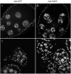
mip120 null egg chambers have a condensed nurse cell DNA phenotype. Stage 7-8 egg chambers from mip120WT control (A) and mip12067-9a-9/Df mutant (B) females were fixed, stained with TO-PRO-3, and imaged by confocal microscopy. Stage 2-5 egg chambers from mip120WT control (C) and mip12067-9a-9/Df mutant (D) females were fixed, stained, and imaged by confocal microscopy.
Fig. 6.
Fig. 6.
Failure of chromosome disassembly in mip120 null ovarian nurse cells. Stage 7-8 egg chambers from mip120WT control (A-C) and mip12067-9a-9/Df mutant (D-F) females were dissected, mounted in halocarbon oil and imaged live by confocal microscopy. Both genotypes also contained transgenes expressing GFP-CID and RFP-HP1. Single channel images of GFP-CID (A,D) and RFP-HP1 (B,E) are shown, as are merged images with GFP-CID (green) and RFP-HP1 (red) (C,F). Autofluorescent yolk material appears in the GFP channel in the mip120WT egg chamber. An outer layer of small follicle cell nuclei surround the large interior nurse cell nuclei.
Fig. 7.
Fig. 7.
Ectopic expression of Rhino does not rescue mip120 null ovarian nurse cells. Ovarioles from mip120WT control (left) and mip12067-9a-9/Df mutant (right) females were dissected, mounted in halocarbon oil and imaged live by confocal microscopy. Both genotypes also contained a nanos-GAL4-VP16 transgene driving germline expression of a UAS-GFP-Rhino transgene. Images show GFP-Rhino present within the nuclei of egg chambers of both genotypes. However, no egg chambers beyond stage 8 are seen in the mip120/Df ovarioles.
Fig. 8.
Fig. 8.
The less conserved N-terminus is required along with the CXC and HCH domains for Mip120 to rescue the mip120 null condensed nurse cell DNA phenotype. Fixed egg chambers from mip120WT (A), mip12067-9a-9/Df (B), mip12067-9a-9/Df; P{mip120}/+ (C), mip12067-9a-9/Df; P{mip120 N-term}/+ (D), mip12067-9a-9/Df; P{mip120 C-term}/+ (E), mip12067-9a-9/Df; P{mip120 CXC}/+ (F), and mip12067-9a-9/Df; P{mip120 HCH}/+ (G) females were stained with TO-PRO-3 and imaged by confocal microscopy.
Fig. 9.
Fig. 9.
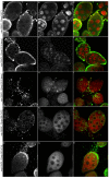
Mip120 CXC and HCH domains are not sufficient for nuclear localization in mip120 null egg chambers. Fixed egg chambers from mip12067-9a-9/Df; P{mip120}/+ (A-C), mip12067-9a-9/Df; P{mip120 N-term}/+ (D-F), mip12067-9a-9/Df; P{mip120 C-term}/+ (G-I), mip12067-9a-9/Df; P{mip120 CXC}/+ (J-L), and mip12067-9a-9/Df; P{mip120 HCH}/+ (M-O) females were fixed and immunostained with anti-Lamin (A,D,G,J,M) to detect the nuclear envelope and anti-Cherry fluorescent protein (B,E,H,K,N) to detect mCherry-Mip120 fusion proteins, then imaged by confocal microscopy. Merged images (C,F,I,L,O) show Lamin in green and Cherry in red.
Fig. 10.
Fig. 10.
A mutant clone homozygous for mip120LL07629 has greatly diminished Mip120 protein levels. hsFLP/+; FRT42B, mip120LL07629/FRT42B, Ubi-GFP-nls females were heat-shocked to induce the site-specific FLP recombinase, generating homozygous GFP-negative clones that were also homozygous for the mutant allele, mip120LL07629. Egg chambers were dissected, fixed, then stained with anti-Mip120 antibodies and TO-PRO-3. Follicle cells on the surface of the egg chamber were imaged by confocal microscopy for: GFP (top left); anti-Mip120 (top right); GFP (green) and anti-Mip120 (red) (bottom left); and TO-PRO-3 (bottom right). The boundary of the GFP-negative mutant clone is indicated by a dashed line. The TO-PRO-3 bright dots within the nuclei represent heterochromatin-dense chromocenters.
Fig. 11.
Fig. 11.
Mip120 is not required in adjacent follicle cells for normal nurse cell morphology. Ovaries were dissected from heat-shocked hsFLP/+; FRT42B, mip120LL07629/FRT42B, Ubi-GFP-nls females as described in Fig. 9, then egg chambers were fixed, dissected, stained with TO-PRO-3 and imaged by confocal microscopy for: GFP (left); TO-PRO-3 (middle); or GFP (green) together with TO-PRO-3 (red) (right). Rightward and upward arrows indicate two egg chambers with large patches of GFP-negative homozygous mip120LL07629 mutant follicle cells adjacent to GFP-positive heterozygous mip120WT/mip120LL07629 nurse cells. Downward arrows indicate an egg chamber with GFP-negative homozygous mip120LL07629 mutant nurse cells surrounded by GFP-positive heterozygous mip120WT/mip120LL07629 follicle cells.
Fig. 12.
Fig. 12.
Levels of bgcn RNA are greatly increased in mip120 null ovaries. Ovaries from mip120WT control (w1118) and mip12067-9a-9/Df(2R)BSC274 mutant females were dissected and qPCR was used to quantitate RNA levels of bam, bgcn, and tut. Two different sets of PCR primers flanking an intron were used for each gene, with the relevant exons indicated as E1, E2, E3, E4. Errors bars indicate RQ min and RQ max values for three technical replicates.

References

    1. Alexandru G., Uhlmann F., Mechtler K., Poupart M.-A. and Nasmyth K. (2001). Phosphorylation of the cohesin subunit Scc1 by Polo/Cdc5 kinase regulates sister chromatid separation in yeast. Cell 105, 459-472. 10.1016/S0092-8674(01)00362-2 - DOI - PubMed
    1. Andrejka L., Wen H., Ashton J., Grant M., Iori K., Wang A., Manak J. R. and Lipsick J. S. (2011). Animal-specific C-terminal domain links myeloblastosis oncoprotein (Myb) to an ancient repressor complex. Proc. Natl. Acad. Sci. USA 108, 17438-17443. 10.1073/pnas.1111855108 - DOI - PMC - PubMed
    1. Bauer C. R., Hartl T. A. and Bosco G. (2012). Condensin II promotes the formation of chromosome territories by inducing axial compaction of polyploid interphase chromosomes. PLoS Genet. 8, e1002873 10.1371/journal.pgen.1002873 - DOI - PMC - PubMed
    1. Beall E. L., Manak J. R., Zhou S., Bell M., Lipsick J. S. and Botchan M. R. (2002). Role for a Drosophila Myb-containing protein complex in site-specific DNA replication. Nature 420, 833-837. 10.1038/nature01228 - DOI - PubMed
    1. Beall E. L., Lewis P. W., Bell M., Rocha M., Jones D. L. and Botchan M. R. (2007). Discovery of tMAC: a Drosophila testis-specific meiotic arrest complex paralogous to Myb-Muv B. Genes Dev. 21, 904-919. 10.1101/gad.1516607 - DOI - PMC - PubMed

LinkOut - more resources