. 2017 Jul;174(3):1476-1491.
doi: 10.1104/pp.17.00193. Epub 2017 May 18.

Structure of eIF4E in Complex with an eIF4G Peptide Supports a Universal Bipartite Binding Mode for Protein Translation

Affiliations

Structure of eIF4E in Complex with an eIF4G Peptide Supports a Universal Bipartite Binding Mode for Protein Translation

Manuel Miras et al. Plant Physiol. 2017 Jul.

Abstract

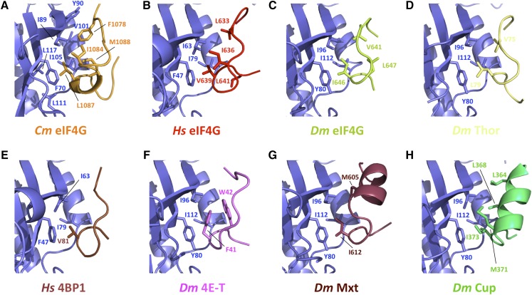
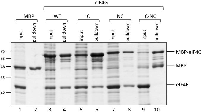
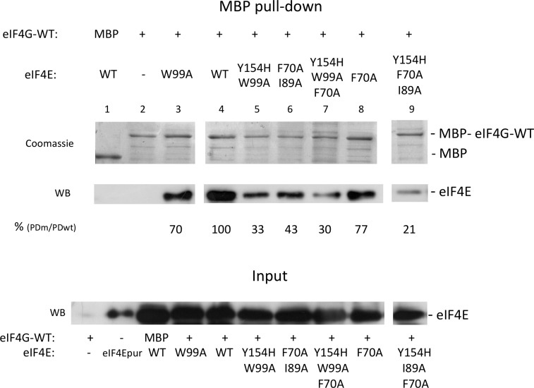
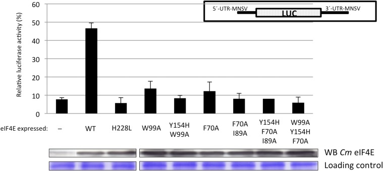
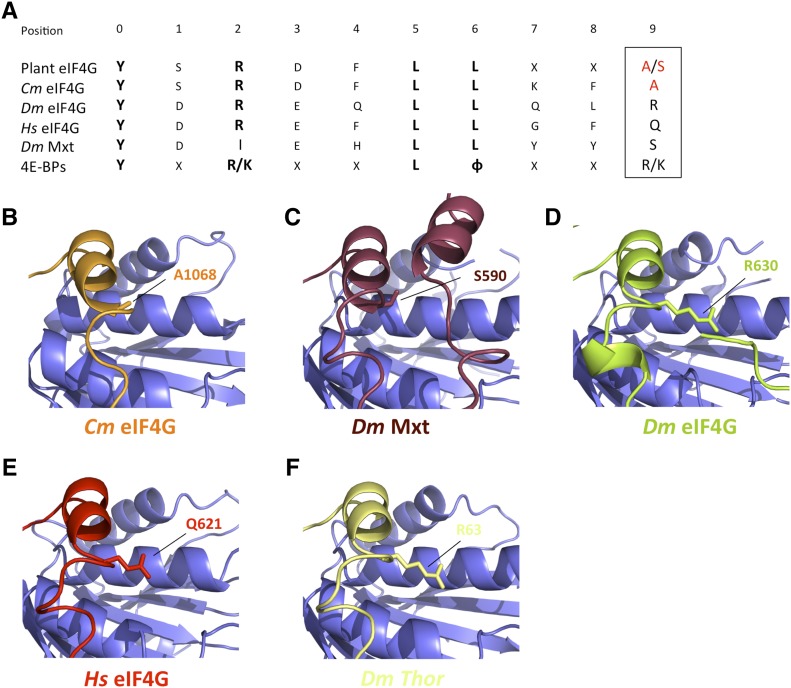
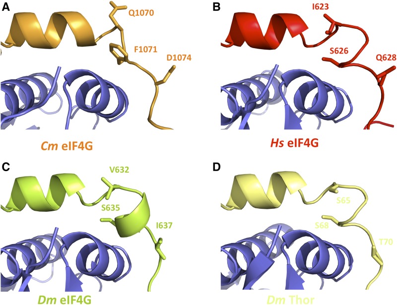
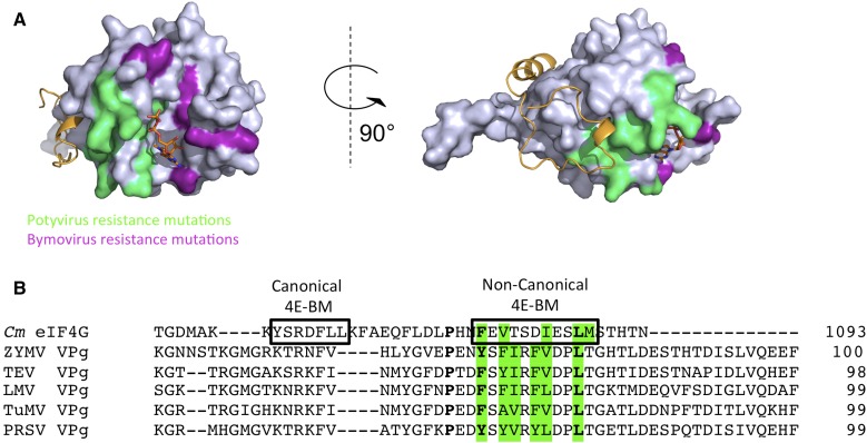
The association-dissociation of the cap-binding protein eukaryotic translation initiation factor 4E (eIF4E) with eIF4G is a key control step in eukaryotic translation. The paradigm on the eIF4E-eIF4G interaction states that eIF4G binds to the dorsal surface of eIF4E through a single canonical alpha-helical motif, while metazoan eIF4E-binding proteins (m4E-BPs) advantageously compete against eIF4G via bimodal interactions involving this canonical motif and a second noncanonical motif of the eIF4E surface. Metazoan eIF4Gs share this extended binding interface with m4E-BPs, with significant implications on the understanding of translation regulation and the design of therapeutic molecules. Here we show the high-resolution structure of melon (Cucumis melo) eIF4E in complex with a melon eIF4G peptide and propose the first eIF4E-eIF4G structural model for plants. Our structural data together with functional analyses demonstrate that plant eIF4G binds to eIF4E through both the canonical and noncanonical motifs, similarly to metazoan eIF4E-eIF4G complexes. As in the case of metazoan eIF4E-eIF4G, this may have very important practical implications, as plant eIF4E-eIF4G is also involved in a significant number of plant diseases. In light of our results, a universal eukaryotic bipartite mode of binding to eIF4E is proposed.


Figures

Figure 1.
Figure 1.
Structure-based sequence alignment of the eIF4E-interacting regions of eIF4G and m4E-BP orthologs. A sequence alignment from superposition of the different eIF4E crystal structures in complex with eIF4E interacting proteins from higher eukaryotes available at the Protein Data Bank (PDB IDs: Cm eIF4G, this work; Hs eIF4G, 5T46; Dm eIF4G, 5T47; Hs 4E-BP1, 4UED; Dm 4E-T, 4UE9; Dm Thor, 4UE8; Dm Mxt, 5ABU) was performed using Chimera (Pettersen et al., 2004). The canonical and NC eIF4E binding motifs (4E-BM) and the elbow loop are framed. The invariant Tyr residue of the canonical motif is typed in bold red and hydrophobic residues conserved in the NC motif are shadowed in blue. Red open circles above the alignment indicate key eIF4G residues within the domains interacting with eIF4E and mutated in this study. The conserved Ser residue in metazoan 4E-BPs is shown in bold blue. The two conserved Ser and Thr residues present in metazoan 4E-BPs implicated in phospho-regulation are colored in orange. Sequences are from Cucumis melo (Cm), Homo sapiens (Hs), and Drosophila melanogaster (Dm).
Figure 2.
Figure 2.
Structure and molecular details of Cm eIF4E unbound or bound to Cm eIF4G1003-1092. A, Ribbon diagram of Cm eIF4E51-235 in complex with the m7GDP cap analog. The m7GDP is located in the cap-binding pocket. Residue W128, in direct interaction with the cap, is marked. B and C, Structure of the Cm eIF4E-eIF4G1003-1092 complex shown as a cartoon and surface rendering, respectively. Cm eIF4G1003-1092 (orange) contains the C and NC eIF4E binding motifs (4E-BM). D, Structural superposition of Cm eIF4E (in gray as cartoon) and Cm eIF4E-eIF4G1003-1092 (in blue as cartoon) binding pocket structures. The formation of a disulfide bridge (shown as sticks in yellow in the structure) promotes the formation of a helix 310 in loop L3 and pulls W128 from m7GDP-interacting position 1 to an apparently “auto-inhibited” position 2 (both labeled in the figure). E, Close-up views of the interaction of the Cm eIF4G C motif (orange) with the dorsal surface of Cm eIF4E (blue) in two orientations. F, Close-up view of the Cm eIF4G linker region (orange) and interactions with Cm eIF4E (blue). G, Close-up view of the NC interaction loop of Cm eIF4G (orange) showing all interactions with the lateral surface of Cm eIF4E (blue). Selected interface residues are shown as blue sticks for Cm eIF4E and as orange sticks for Cm eIF4G. In E to G, residues implicated in the interactions between Cm eIF4E and Cm eIF4G1003-1092 are shown as sticks.
Figure 3.
Figure 3.
Structures of the eIF4E lateral hydrophobic pocket interacting with eIF4G and m4E-BP peptides. Hydrophobicity of residues in all eIF4E-interacting domains is conserved. Close-up views of eIF4E hydrophobic patch and residues involved in the binding of Cm eIF4E and eIF4G1003-1092 (A), Hs eIF4E and eIF4G592-653 (B; PDB ID: 5T46; Grüner et al., 2016), Dm eIF4E and eIF4G601-660 (C; PDB ID: 5T47; Grüner et al., 2016), Dm eIF4E and m4E-BP Thor50-83 (D; PDB ID: 4UE8; Peter et al., 2015a), Hs eIF4E and m4E-BP 4E-BP150-83 (E; PBD ID: 4UED; Peter et al., 2015a), Dm eIF4E and m4E-BP 4E-T9-44 (F; PBD ID: 4UE9; Peter et al., 2015a), Dm eIF4E and m4E-BP Mxt577-640 (G; PBD ID: 5ABV; Peter et al, 2015b), and Dm eIF4E and m4E-BP Cup325-376 (H; PBD ID: 4AXG; Kinkelin et al., 2012).
Figure 4.
Figure 4.
In vitro interaction of Cm eIF4E and Cm eIF4G1003-1092. MBP pull-down assay showing the interaction of Cm eIF4E (wild type, full length) and MBP-Cm eIF4G1003-1092 (either wild type or mutated in C, NC, or C-NC). Bacterial lysates expressing MBP-tagged Cm eIF4G1003-1092 (wild type and mutants) were incubated with purified Cm eIF4E 1 μm in a volume of 300 μL. Of these, 15 μL (5%; input) was analyzed by SDS-PAGE followed by Coomassie Brilliant Blue staining. Bound proteins were eluted in 60 μL, and 10 μL (16%; pulldown) was loaded on the gel. MBP served as a negative control (lanes 1 and 2).
Figure 5.
Figure 5.
Binding affinity of Cm eIF4E and Cm eIF4G1003-1092. MBP pull-down assays showing the interaction of purified Cm eIF4E (wild type, full length; increasing concentrations, 0.5–3 μm) and MBP-tagged Cm eIF4G1003-1092 (wild type or mutant in bacterial lysates at a fixed concentration). A, Pull-down with Cm eIF4G1003-1092-WT. B, Pull-down with MBP used as a negative control. C to E, Pull-down with MBP-eIF4G1003-1092 mutants in C, NC, and C-NC domains. EIF4G lysates (wild type and mutants) were incubated with purified Cm eIF4E at concentrations of 0.5, 1, 2, and 3 μm (corresponding to lanes 1, 2, 3, and 4 of each gel, respectively) in a volume of 300 μL. The input samples (lanes 1 to 4) and pull-down fractions (lanes 5 to 8) were analyzed in a 12% SDS-PAGE followed by Coomassie Brilliant Blue staining, loading in gels 5% and 16% of the total volumes, respectively. F, Graph representing the specific binding of Cm eIF4E with Cm eIF4G1003-1092 (wild type and mutants), plotted as a function of Cm eIF4E concentration. Data points in the binding curves represent means calculated from data points of two different experiments as in A, C, and D. Each data was fitted using a one-site binding model.
Figure 6.
Figure 6.
Recovery of Cm eIF4E (wild type and mutants) in pull-down assays with MBP-Cm eIF4G1003-1092. Pull-down assays showing the interaction of wild-type and mutant Cm eIF4E with MBP-Cm eIF4G-WT. A fixed volume of a lysate from bacteria expressing Cm eIF4G1003-1092-WT with a N-terminal MBP tag was incubated with Cm eIF4E bacterial lysates (wild type and mutants) in a volume of 300 μL. Middle, Pulled-down wild-type and mutant Cm eIF4Es as determined by Western blot (WB) using a Cm eIF4E-specific antibody. Top, The same gel was stained with Coomassie Brilliant Blue to compare the amount of eluted MBP-tagged Cm eIF4G-WT in each experiment (Coomassie). Bottom, The amount of Cm eIF4E present in the input visualized by western blot. Bands from WB were quantified and a binding was estimated using the band intensity ratio between sample Cm eIF4E-WT (input, lane 5) and Cm eIF4E-WT (MBP pull-down, lane 4) as 100% reference value. The interacting factors in each experiment are shown below the gels: pull-down proteins, either MBP or MBP-Cm eIF4G, and interacting Cm eIF4E proteins (wild type or mutants). Input and pull-down correspond to a 1% of the incubation volume and 5% of the elution volumes, respectively.
Figure 7.
Figure 7.
Effect of substitutions in Cm eIF4E on cap-independent translation efficiency. Relative luciferase activity (%) obtained with a reporter RNA that depends on Cm eIF4E for cap-independent translation (see inset in the right upper part of figure) melon protoplasts. Protoplasts carried Cm eIF4EH228L, which is unable to efficiently complement cap-independent translation of the reporter RNA. Protoplasts were transiently transformed to express different Cm eIF4E variants (wild type, which is able to complement cap-independent translation of the reporter; Miras et al., 2017b; and mutants). For normalization of measurements in different protoplast preparations, luciferase activity obtained with a Cm eIF4E independently translated reporter RNA construct 5′-UTR-luc-3′-UTR was set to 100% for each protoplast preparation. The transiently expressed Cm eIF4E mutants are indicated below each bar: -, silencing suppressor P19 alone; WT, wild type; engineered Cm eIF4E mutations, H228L, W99A and Y154H-W99A (in residues in the dorsal surface proposed to be involved in canonical Cm eIF4G interaction; Miras et al., 2017b), and F70A and F70A-I89A (in residues in the lateral surface proposed to be involved in NC Cm eIF4G interaction); and mutations in both surfaces Y154H-F70A-I89A and W99A-Y154H-F70A. Error bars are ±sd. Panels below the graph show expression of each Cm eIF4E mutant in melon cotyledons visualized by western blot using antibodies against Cm eIF4E. Bottom, Loading as visualized by Coomassie Brilliant Blue staining.
Figure 8.
Figure 8.
Structural comparison of Cm eIF4G, metazoan eIF4Gs, and 4E-BP linker regions. A, Extended consensus sequence for the plant eIF4G canonical eIF4E binding domain from all available databank sequences (Patrick and Browning, 2012) and Dm Mxt and consensus m4E-BPs. Invariant residues are typed in bold, and plant-specific Ala or Ser residues in position 9 (boxed) are red colored. B to F, Close-up views of the canonical helices and the linker region of Cm eIF4G (B), Dm Mxt (C), Dm eIF4G (D), Hs eIF4G (E), and Dm Thor (as m4E-BP model; F) bound to the dorsal surface of eIF4E (PDB IDs: 5T46, 5T47, 5ABV, 4UE8; Peter et al., 2015a, 2015b; Grüner et al., 2016). Residues of the canonical helix in position 9 are shown as sticks.
Figure 9.
Figure 9.
Structural comparison of the elbow loops of Cm eIF4G, metazoan eIF4Gs, and 4E-BP. A to D, Close-up views of the canonical helices and elbow loops of Cm eIF4G (A), Hs eIF4G (B), Dm eIF4G (C), and Dm Thor (as a m4E-BP model; D) bound to the dorsal surface of eIF4E (PDB IDs: 5T46, 5T47, 4UE8; Peter et al., 2015a; Grüner et al., 2016). Selected residues mediating phosphorylation and their analogs are shown as sticks.
Figure 10.
Figure 10.
Plant virus resistance mutations modeled on the melon eIF4E-eIF4G1003-1092 complex. A, The residues implicated in plant potyvirus and bymovirus resistance are shaded in green and magenta, respectively (Gao et al., 2004; Kang et al., 2005; Kanyuka et al., 2005; Ruffel et al., 2005; Stein et al., 2005; Nicaise et al., 2007; German-Retana et al., 2008; Ashby et al., 2011). The Cm eIF4E molecule is shown as a surface rendering and Cm eIF4G1003-1092 is shown as a cartoon representation, both colored in gray and orange, respectively. The m7GDP cap-analog is located in the cap-binding pocket of Cm eIF4E. B, Sequence alignment of Cm eIF4G (residues 1052–1093) and potyviral VPgs (ZYMV NP477522.1; TEV NP734204.1; LMV KF285932.1; TuMV NP734219.1; PRSV AEC04846.1). C and NC motifs are boxed in the Cm eIF4G sequence. Conserved residues are typed in bold and hydrophobic residues located in the putative NC motif are green-colored.

Similar articles

Cited by

References

    1. Adams PD, Grosse-Kunstleve RW, Hung LW, Ioerger TR, McCoy AJ, Moriarty NW, Read RJ, Sacchettini JC, Sauter NK, Terwilliger TC (2002) PHENIX: building new software for automated crystallographic structure determination. Acta Crystallogr D Biol Crystallogr 58: 1948–1954 - PubMed
    1. Aitken CE, Lorsch JR (2012) A mechanistic overview of translation initiation in eukaryotes. Nat Struct Mol Biol 19: 568–576 - PubMed
    1. Altmann M, Müller PP, Pelletier J, Sonenberg N, Trachsel H (1989) A mammalian translation initiation factor can substitute for its yeast homologue in vivo. J Biol Chem 264: 12145–12147 - PubMed
    1. Ashby JA, Stevenson CEM, Jarvis GE, Lawson DM, Maule AJ (2011) Structure-based mutational analysis of eIF4E in relation to sbm1 resistance to pea seed-borne mosaic virus in pea. PLoS One 6: e15873. - PMC - PubMed
    1. Bah A, Vernon RM, Siddiqui Z, Krzeminski M, Muhandiram R, Zhao C, Sonenberg N, Kay LE, Forman-Kay JD (2015) Folding of an intrinsically disordered protein by phosphorylation as a regulatory switch. Nature 519: 106–109 - PubMed

MeSH terms

Substances

LinkOut - more resources