

Structure-function analysis of human sucrase-isomaltase identifies key residues required for catalytic activity
- PMID: 28522605
- PMCID: PMC5491789
- DOI: 10.1074/jbc.M117.791939
Structure-function analysis of human sucrase-isomaltase identifies key residues required for catalytic activity
Abstract
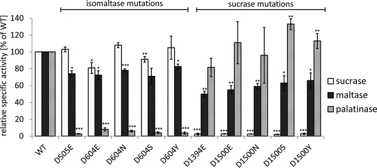
Sucrase-isomaltase (SI) is an intestinal membrane-associated α-glucosidase that breaks down di- and oligosaccharides to absorbable monosaccharides. SI has two homologous functional subunits (sucrase and isomaltase) that both belong to the glycoside hydrolase family 31 (GH31) and differ in substrate specificity. All GH31 enzymes share a consensus sequence harboring an aspartic acid residue as a catalytic nucleophile. Moreover, crystallographic structural analysis of isomaltase predicts that another aspartic acid residue functions as a proton donor in hydrolysis. Here, we mutagenized the predicted proton donor residues and the nucleophilic catalyst residues in each SI subunit. We expressed these SI variants in COS-1 cells and analyzed their structural, transport, and functional characteristics. All of the mutants revealed expression levels and maturation rates comparable with those of the wild-type species and the corresponding nonmutated subunits were functionally active. Thereby we determined rate and substrate specificity for each single subunit without influence from the other subunit. This approach provides a model for functional analysis of the single subunits within a multidomain protein, achieved without the necessity to express the individual subunits separately. Of note, we also found that glucose product inhibition regulates the activities of both SI subunits. We experimentally confirmed the catalytic function of the predicted proton donor residues, and sequence analysis suggested that these residues are located in a consensus region in many GH31 family members. In summary, these findings reveal the kinetic features specific for each human SI subunit and demonstrate that the activities of these subunits are regulated via product inhibition.
Keywords: Glycoside hydrolase family 31; carbohydrate metabolism; enzyme kinetics; glycosidase; product inhibition; site-directed mutagenesis; starch digestion; substrate specificity; sucrase-isomaltase.
© 2017 by The American Society for Biochemistry and Molecular Biology, Inc.
Conflict of interest statement
The authors declare that they have no conflicts of interest with the contents of this article.
Figures


Similar articles
-
Phylogenetic analysis reveals key residues in substrate hydrolysis in the isomaltase domain of sucrase-isomaltase and its role in starch digestion.Biochim Biophys Acta Gen Subj. 2019 Sep;1863(9):1410-1416. doi: 10.1016/j.bbagen.2019.06.011. Epub 2019 Jun 27. Biochim Biophys Acta Gen Subj. 2019. PMID: 31254546
-
Sucrase is an intramolecular chaperone located at the C-terminal end of the sucrase-isomaltase enzyme complex.J Biol Chem. 2002 Aug 30;277(35):32141-8. doi: 10.1074/jbc.M204116200. Epub 2002 Jun 7. J Biol Chem. 2002. PMID: 12055199
-
Molecular pathogenicity of novel sucrase-isomaltase mutations found in congenital sucrase-isomaltase deficiency patients.Biochim Biophys Acta Mol Basis Dis. 2017 Mar;1863(3):817-826. doi: 10.1016/j.bbadis.2016.12.017. Epub 2017 Jan 3. Biochim Biophys Acta Mol Basis Dis. 2017. PMID: 28062276
-
Structural Studies of the Intestinal α-Glucosidases, Maltase-glucoamylase and Sucrase-isomaltase.J Pediatr Gastroenterol Nutr. 2018 Jun;66 Suppl 3:S11-S13. doi: 10.1097/MPG.0000000000001953. J Pediatr Gastroenterol Nutr. 2018. PMID: 29762369 Review.
-
Naturally occurring sulfonium-ion glucosidase inhibitors and their derivatives: a promising class of potential antidiabetic agents.Acc Chem Res. 2014 Jan 21;47(1):211-25. doi: 10.1021/ar400132g. Epub 2013 Aug 22. Acc Chem Res. 2014. PMID: 23964564 Review.
Cited by
-
The Hippo Pathway Effector YAP1 Regulates Intestinal Epithelial Cell Differentiation.Cells. 2020 Aug 13;9(8):1895. doi: 10.3390/cells9081895. Cells. 2020. PMID: 32823612 Free PMC article.
-
Theoretical Studies for the Discovery of Potential Sucrase-Isomaltase Inhibitors from Maize Silk Phytochemicals: An Approach to Treatment of Type 2 Diabetes.Molecules. 2023 Sep 23;28(19):6778. doi: 10.3390/molecules28196778. Molecules. 2023. PMID: 37836621 Free PMC article.
-
The Role of Metal Oxide Nanoparticles, Escherichia coli, and Lactobacillus rhamnosus on Small Intestinal Enzyme Activity.Environ Sci Nano. 2020 Dec 1;7(12):3940-3964. doi: 10.1039/d0en01001d. Epub 2020 Nov 9. Environ Sci Nano. 2020. PMID: 33815806 Free PMC article.
-
Different Trafficking Phenotypes of Niemann-Pick C1 Gene Mutations Correlate with Various Alterations in Lipid Storage, Membrane Composition and Miglustat Amenability.Int J Mol Sci. 2020 Mar 19;21(6):2101. doi: 10.3390/ijms21062101. Int J Mol Sci. 2020. PMID: 32204338 Free PMC article.
-
Bioactive Peptides from Germinated Soybean with Anti-Diabetic Potential by Inhibition of Dipeptidyl Peptidase-IV, α-Amylase, and α-Glucosidase Enzymes.Int J Mol Sci. 2018 Sep 22;19(10):2883. doi: 10.3390/ijms19102883. Int J Mol Sci. 2018. PMID: 30249015 Free PMC article.
References
-
- Naim H. Y., Sterchi E. E., and Lentze M. J. (1988) Biosynthesis of the human sucrase-isomaltase complex: differential O-glycosylation of the sucrase subunit correlates with its position within the enzyme complex. J. Biol. Chem. 263, 7242–7253 - PubMed
-
- Treem W. R. (1995) Congenital sucrase-isomaltase deficiency. J. Pediatr. Gastroenterol. Nutr. 21, 1–14 - PubMed
-
- Hunziker W., Spiess M., Semenza G., and Lodish H. F. (1986) The sucrase-isomaltase complex: primary structure, membrane-orientation, and evolution of a stalked, intrinsic brush border protein. Cell 46, 227–234 - PubMed
-
- Sander P., Alfalah M., Keiser M., Korponay-Szabo I., Kovacs J. B., Leeb T., and Naim H. Y. (2006) Novel mutations in the human sucrase-isomaltase gene (SI) that cause congenital carbohydrate malabsorption. Hum. Mutat. 27, 119 - PubMed
MeSH terms
Substances
Associated data
- Actions
- Actions
LinkOut - more resources
Full Text Sources
Other Literature Sources
