. 2017 May 18;7(1):2102.
doi: 10.1038/s41598-017-02055-x.

Selection and characterization of a DNA aptamer inhibiting coagulation factor XIa

Affiliations

Selection and characterization of a DNA aptamer inhibiting coagulation factor XIa

David A Donkor et al. Sci Rep. .

Abstract

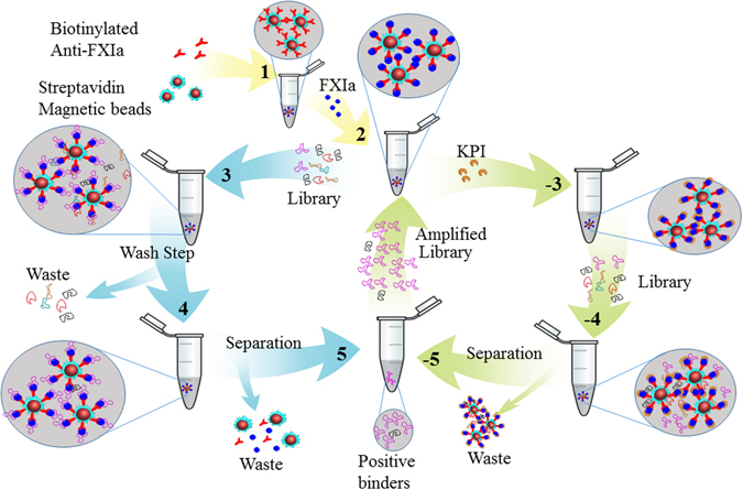
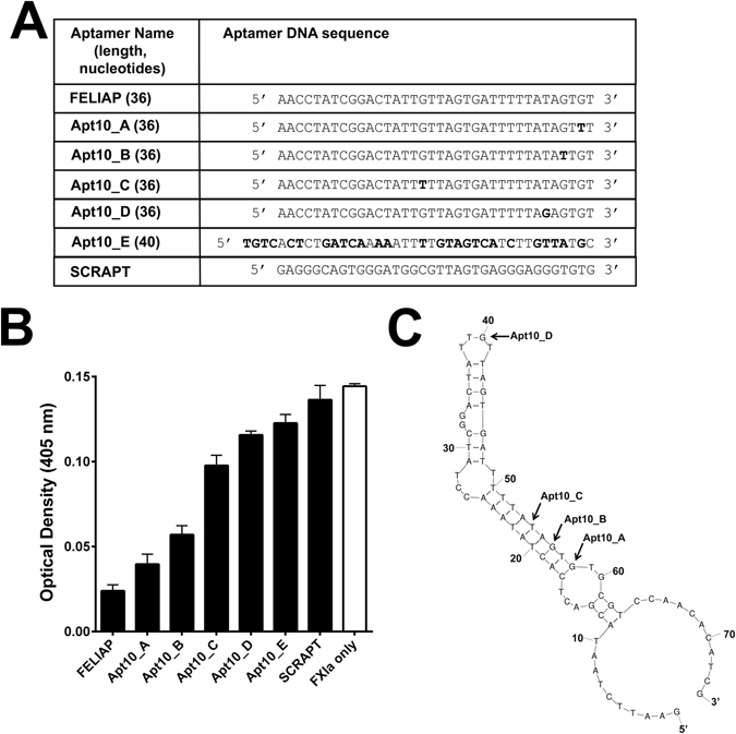
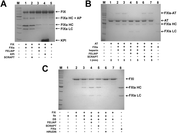
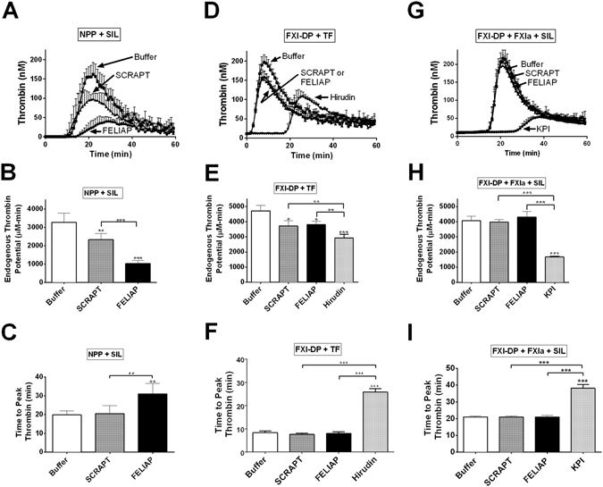
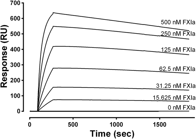
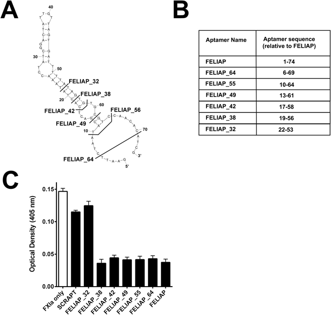
Factor XIa (FXIa) is a serine protease that catalyzes the activation of Factor IX (FIX) in the blood coagulation cascade. FXIa and its precursor FXI are emergent therapeutic targets for the development of safer anticoagulant agents. Here, we sought a novel DNA-based agent to inhibit FXIa. Towards this goal, an 80 base, single-stranded DNA aptamer library (containing a 40 base randomized core) was screened for FXIa-binding candidates, using ten rounds of positive and negative selection. After selection, 6 of 89 different sequences inhibited FXIa-mediated chromogenic substrate S2366 cleavage. The most active anti-FXIa aptamer had a hypervariable central sequence 5'-AACCTATCGGACTATTGTTAGTGATTTTTATAGTGT-3' and was designated Factor ELeven Inhibitory APtamer (FELIAP). FELIAP, but not a scrambled aptamer control (SCRAPT), competitively inhibited FXIa-catalyzed S2366 cleavage, FIX activation, and complex formation with antithrombin. No effect of FELIAP on FXI activation was observed. FELIAP inhibited plasma clotting and thrombin generation assays to a significantly greater extent than SCRAPT. Immobilized FELIAP bound FXIa with strong affinity and an equilibrium binding constant (KD) in the low nanomolar range determined using surface plasmon resonance. FELIAP is the first FXIa-inhibitory aptamer to be described and constitutes a lead compound to develop related aptamers for in vivo use.


Conflict of interest statement

The authors declare that they have no competing interests.

Figures

Figure 1
Figure 1
Schematic representation of aptamer library screening strategy. For rounds in which only positive selection was employed, biotinylated anti-FXIa antibodies and streptavidin-coated magnetic beads (1) were combined with FXIa (2) and the original aptamer library (3). Aptamer-FXIa-antibody-bead complexes were then concentrated magnetically and washed (4) prior to separation and recovery of selected aptamers via phenol/chloroform/isoamyl alcohol extraction and ethanol precipitation (5). Following asymmetric PCR to amplify the aptamer pool, steps 1–5 were either repeated directly or components from 1 and 2 were combined with the FXIa-active site inhibitor KPI (−3) for negative selection, magnetic concentration and washing (−4), separation (−5) and amplification, to complete rounds in which both positive and negative selection were employed.
Figure 2
Figure 2
Characterization of Round 10 aptamers as inhibitors of FXIa-mediated amidolysis. (A) Means ± SEM (n = 3) of colour generation following FXIa-mediated amidolysis of chromogenic substrate S2366, in the presence (black bars) of Round 10 aptamers identified on the x axis or a scrambled negative control sequence (SCRAPT), or in the absence of added DNA molecules (white bar). (B) Mfold-generated predicted secondary structure of FELIAP. The positions of base substitutions between FELIAP and other Round 10-selected aptamers are indicated by arrows. (C) DNA sequence of Round 10 aptamers and SCRAPT. The sequence corresponding to the original 40 nucleotide hypervariable core of the aptamer library is shown, with base substitutions relative to FELIAP in bold.
Figure 3
Figure 3
Kinetic characterization of FELIAP inhibition of FXIa-mediated amidolysis. (A) Means ± SEM (n = 3) of reaction velocity for FXIa-mediated amidolysis of S2366 versus aptamer concentration (FELIAP, circles; SCRAPT, squares). (B) Means ± SEM (n = 3) of reaction velocity versus S2366 concentration in the presence of 0, 10, or 20 µM FELIAP. (C) Lineweaver-Burke transformation of data in (B). (D) Means ± SEM (n = 7) of modified APTT assays in which FELIAP or SCRAPT were pre-incubated with FXIa, at concentrations given on the x axis, prior to dilution into recalcified FXI-deficient plasma.
Figure 4
Figure 4
Effects of FELIAP on reactions of FXIa and FXI with macromolecular substrates. (A) Coomassie-stained reduced SDS-polyacrylamide gel. FIX was reacted with FXIa in the presence (+) or absence (−) of SCRAPT (lane 2), FELIAP (lane 3) or KPI (lane 4). Arrows and labels, at right, identify the position of FIX, forms of FIXa (including HC, heavy chain, HC + AP, heavy chain + activation peptide intermediate, LC, light chain) and KPI. M, markers (kDa), at left: 220; 160; 120; 100; 90; 80; 70; 60; 50; 40; 30; 25; 20; 15. (B) As in A, except antithrombin (AT, lane 1) was reacted with FXIa (lane 8) in the presence of heparin for 1 (lane 2) or 5 (lane 3) minutes with FELIAP (lanes 4 and 5) or SCRAPT (lanes 6 and 7) or no DNA addition (lanes 2 and 3). Arrows and labels identify the position of AT, FXIa-AT covalent complex, FXIa heavy chain (HC) and light chain (LC). M, markers (kDa), at left: 220; 160; 120; 100; 90; 80; 70; 60; 50; 40; 30; 25; 20 (C). As in A and B except FXI and thrombin (IIa, lane 1) were reacted in the absence (lane 2) or presence of dextran sulphate (DS, lanes 3–7) with the addition of FELIAP (lane 4) or SCRAPT (lane 5) or the thrombin inhibitor hirudin (lane 7). Lane 8, purified FXIa. M, markers (kDa), at left: 220; 160; 120; 100; 90; 80; 70; 60; 50; 40; 30; 25; 20; 15.
Figure 5
Figure 5
Inhibition of thrombin generation by FELIAP. Thrombin generation assays (TGA) were conducted in 3 different ways: in recalcified human normal pooled plasma (NPP) using micronized silica (SIL) for contact activation as the initiator (NPP + SIL, panels A-C); in FXI-depleted plasma (FXI-DP) using tissue factor for extrinsic pathway activation as the initiator (FXI-DP + TF, panels D-F); and following 0.25 nM FXIa pre-incubation with Buffer, 1 µM SCRAPT, or 1 µM FELIAP, in FXI-DP activated with micronized silica (FXI-DP + FXIa + SIL). Thrombin concentration was determined fluorescently every minute for 60 minutes in each case. (A) TGA progress curves (mean ± SD (n = 6) with addition of agents (Buffer, 30 μM FELIAP, or 30 μM SCRAPT) as indicated by labels and arrows. Upwards error bars are shown. (D) As in A, but n = 5, and with additional assays including 0.2 µM recombinant hirudin variant 3 (Hirudin). (G) FELIAP, SCRAPT or KPI was pre-incubated with FXIa (1 µM: 0.25 mM inhibitor: enzyme ratio) and then diluted into FXI-DP activated with micronized silica for TGA. Progress curves (mean ± SD (n = 5)) are shown. Each set of TGA progress curves (A,D,G) are quantified with respect to endogenous thrombin potential (the area under the thrombogram curve) (B,E,H) and time to peak thrombin (C,F,I) in panel columns. Bar graphs are derived from analysis of individual thrombogram curves corresponding to plasma supplementation with Buffer (white), SCRAPT (grey), FELIAP (black), or, in some reactions KPI or Hirudin (light grey). Symbols (above the error bars) indicate statistically significant differences from Buffer reactions, while symbols above the horizontal bar indicate statistically significant differences between SCRAPT- and FELIAP-supplemented and other reactions: p < 0.001, *; *p < 0.001, ***.
Figure 6
Figure 6
Binding of FELIAP to immobilized FXIa. An SPR sensogram showing the interactions between 3′biotinylated FELIAP immobilized on a streptavidin-coated chip and FXIa at 25 °C at FXIa concentrations given above each progress curves. Association was allowed to proceed for 180 seconds followed by 1800 seconds of dissociation. All curves were corrected for non-specific effects by signal subtraction using a reference cell containing 3′biotinylated SCRAPT immobilized on a streptavidin-coated chip. A single dilution series of curves representative of a total of 3 others is shown.
Figure 7
Figure 7
Characterization of truncated derivatives of FELIAP as inhibitors of FXIa-mediated amidolysis. (A) Extent of truncation analysis. Sequences to the left of bolded lines crossing the Mfold-generated predicted secondary structure of FELIAP are identified as FELIAP_X, where X = the length of the truncated aptamer (32, 38, 42, 49, 55, 64, or 74 for full-length FELIAP. (B) Tabular depiction of truncated aptamers relative to FELIAP; for instance, FELIAP_32 (relative sequence 22–53) is identical to FELIAP except for the deletion of 21 nucleotides from the 5′end and 21 nucleotides from the 3′ end of FELIAP. (C) Means ± SEM (n = 3) of colour generation following FXIa-mediated amidolysis of chromogenic substrate S2366, in the presence (black bars) of SCRAPT, FELIAP, or truncated derivatives of FELIAP identified in panels A and B, or in the absence of of added DNA molecules (white bar).

Similar articles

Cited by

References

    1. Gale AJ. Continuing education course #2: current understanding of hemostasis. Toxicol Pathol. 2011;39:273–280. doi: 10.1177/0192623310389474. - DOI - PMC - PubMed
    1. Furie B, Furie BC. Mechanisms of thrombus formation. N Engl J Med. 2008;359:938–949. doi: 10.1056/NEJMra0801082. - DOI - PubMed
    1. Mackman N. New insights into the mechanisms of venous thrombosis. J Clin Invest. 2012;122:2331–2336. doi: 10.1172/JCI60229. - DOI - PMC - PubMed
    1. Lozano R, et al. Global and regional mortality from 235 causes of death for 20 age groups in 1990 and 2010: a systematic analysis for the Global Burden of Disease Study 2010. Lancet. 2012;380:2095–2128. doi: 10.1016/S0140-6736(12)61728-0. - DOI - PMC - PubMed
    1. Hirsh J. Oral anticoagulant drugs. N Engl J Med. 1991;324:1865–1875. doi: 10.1056/NEJM199106273242606. - DOI - PubMed

Publication types

MeSH terms

LinkOut - more resources