. 2017 May 18;7(1):2108.
doi: 10.1038/s41598-017-01108-5.

Near-Infrared Heptamethine Cyanine Based Iron Oxide Nanoparticles for Tumor Targeted Multimodal Imaging and Photothermal Therapy

Affiliations

Near-Infrared Heptamethine Cyanine Based Iron Oxide Nanoparticles for Tumor Targeted Multimodal Imaging and Photothermal Therapy

Sejy Lee et al. Sci Rep. .

Abstract

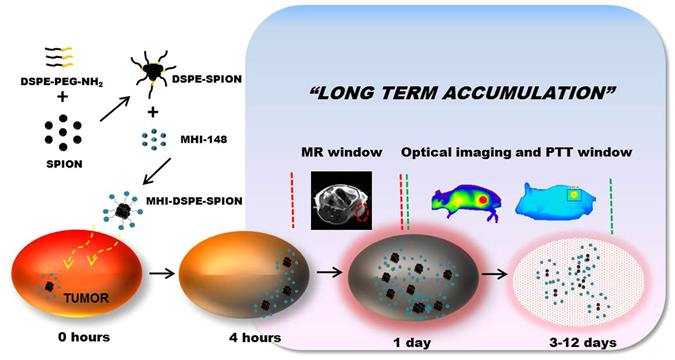
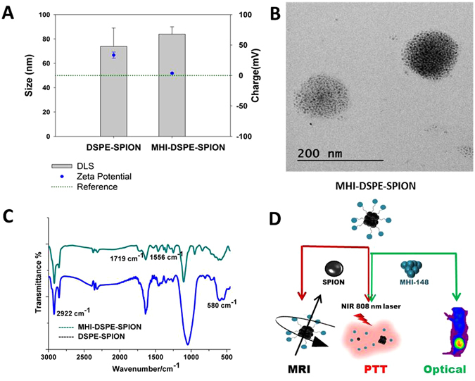
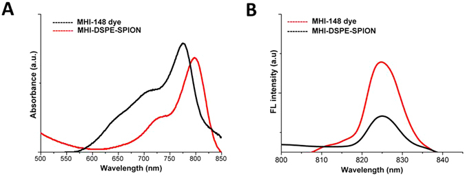
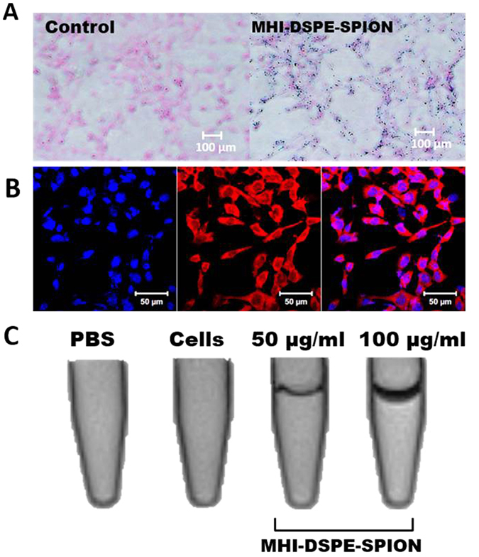
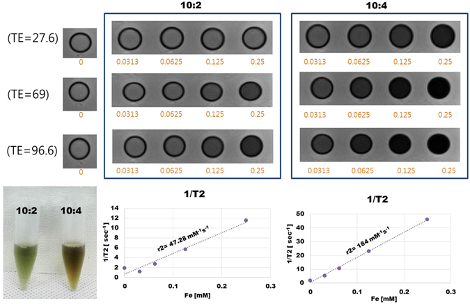
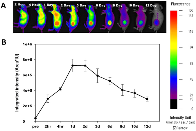
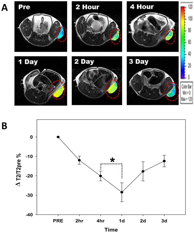
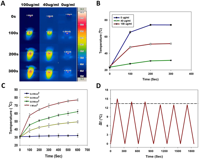
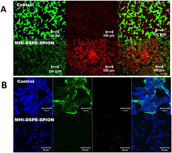
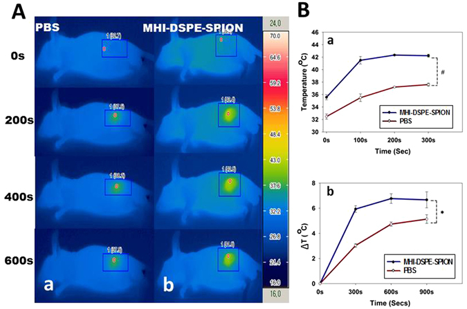
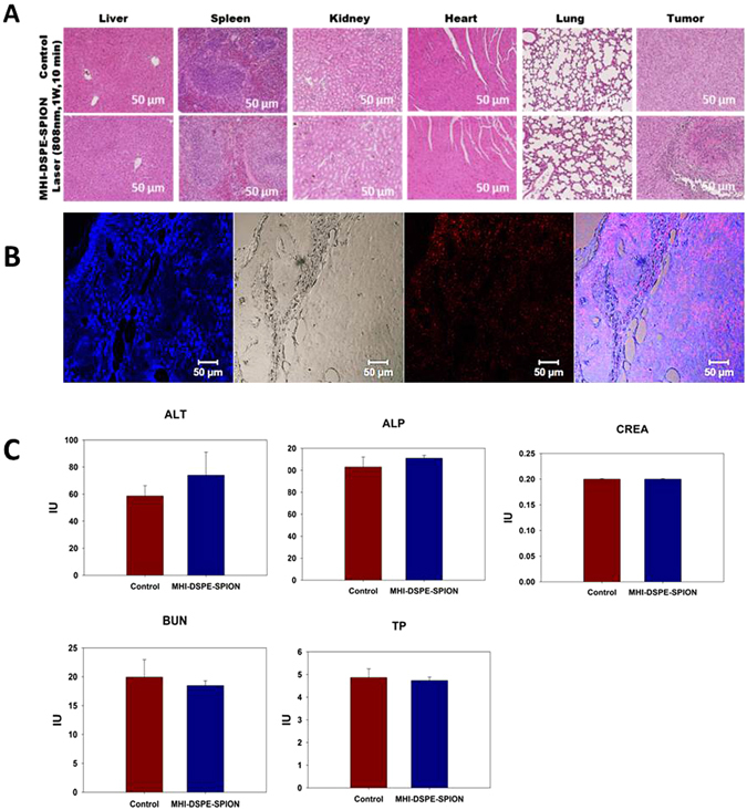
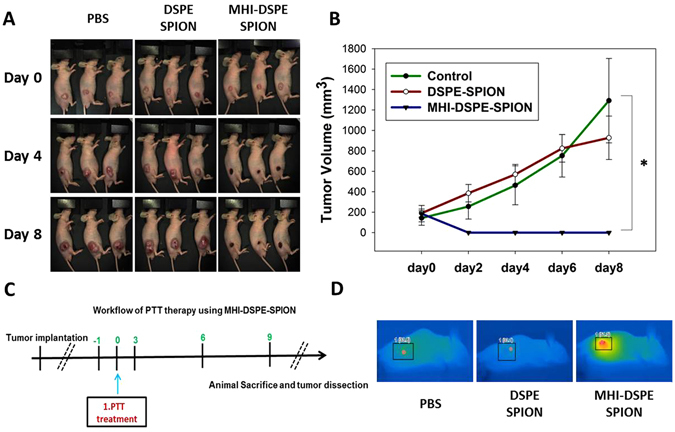
Near-infrared fluorescent (NIRF) imaging modality holds great promise for tumor detection and offers several advantages of bioimaging, such as high tissue penetration with less background scattering. The disadvantage of NIRF bioimaging is that it has very low spatial resolution. Thus, the combination of NIRF with magnetic resonance imaging (MRI) is a good option because MRI can provide anatomical information with a higher resolution. Heptamethine cyanine dye (MHI-148) has been reported to have tumor-targeting capability which was used here as the NIRF agent. DSPE-SPION nanoparticles were synthesized by the solvent hydration method and conjugated with MHI-148 dye to form a MRI/NIRF dual imaging probe. The size and charge of the MHI-DSPE-SPION were found to be about 84 ± 6 nm and 3.7 mV by DLS & Zeta Potential analysis. In vivo MRI of the SCC7 tumor showed an enhanced accumulation of MHI-DSPE-SPION, peaking at day 1, compared to 4 hrs with the control DSPE-SPION. An in vivo photothermal tumor reduction study was done on the SCC7 tumor of BALB/c nude mice. Tumor reduction study showed complete tumor removal after 8 days. In conclusion, MHI-DSPE-SPION can be used as a cancer theranostics material because it provides MRI-optical imaging capabilities and the photothermal therapy (PTT) effect.


Conflict of interest statement

The authors declare that they have no competing interests.

Figures

Figure 1
Figure 1
Schematic illustration showing the overall concept of the present study. MHI-DSPE-SPION designed for MRI application by using the magnetism of the nanoparticle core of SPION (black) in addition to optical imaging along with PTT by MHI-148 ligands (green) of the nanoparticle shell (DSPE-PEG).
Figure 2
Figure 2
Physicochemical properties of MHI-DSPE-SPION and DSPE-SPION. (A) Surface charge and hydrodynamic size, as measured by zeta potential and DLS analysis, respectively. (B) MHI-DSPE-SPION morphology, as observed by TEM (C) FT-IR was done in the spectral range between 800 and 3000 cm−1 for DSPE-SPION and MHI-DSPE-SPION to analyze the bonding between DSPE-SPION and the MHI-148 dye. (D) Illustration describing the role of SPION and MHI-148.
Figure 3
Figure 3
Ultraviolet–visible absorbance and fluorescence of free (A) MHI-148 dye and (B) MHI-DSPE-SPION nanoparticles.
Figure 4
Figure 4
Cytotoxicity of MHI-DSPE-SPION analysed using NIH3T3 cells, as quantified by MTS assay. Mean cell viability of NIH3T3 was used for MTS assay in quadruplicate samples ± SD.
Figure 5
Figure 5
Uptake of MHI-DSPE-SPION in SCC7 cells. The dosage was 50 µg/mL, and the incubation time was 2 hrs. (A) Prussian blue staining images show significant uptake of MHI-DSPE-SPION in SCC7 cells compared to the control. (B) CLSM image showing MHI-DSPE-SPION (red) and nuclei stained for 40, 6-diamidino-2-phenylindole (DAPI), and a merged image made from NIR and DAPI images. (C) In vitro phantom tube MRI of SCC7 cells shows a dark band within the tube with the intracellular uptake of MHI-DSPE-SPION. A thicker band was shown for 100 µg/mL dosage, which proves concentration based uptake efficiency increase in SCC7 cells.
Figure 6
Figure 6
Relaxivity (r2 value) measurements of MHI-DSPE-SPION by linear correlation between the relaxation rate, 1/T2, and the concentration of iron for ratios of 10:2 and 10:4.
Figure 7
Figure 7
NIR fluorescence and biodistribution for MHI-DSPE-SPION injected BALB/c nude mice in vivo. NIR images of mice bearing the SCC7 tumor, captured within 2 hrs after i.v. injection of MHI-DSPE-SPION at a dose 10 mg[Fe]/kg. The mice were subjected to fluorescence imaging (NIR) using the FOBI imaging system. (A) In vivo NIR fluorescence of BALB/c nude mice, measured at different times. (B) Quantitative analysis of tumor fluorescence intensity (ROI), measured as integrated intensity (Area*IU).
Figure 8
Figure 8
In vivo MRI study. (A) In vivo T2-weighted MRI of MHI-DSPE-SPION. (B) Signal change of the SCC7 tumor in T2-weighted MRI, measured at different time points (pre, 2 hrs, 4 hrs, 1d, 2d, 3d) for 10 mg[Fe]/kg. MHI-DSPE-SPION (in PBS) was injected through the tail vein. *P < 0.05 relative to the pre-injection T2 contrast (n = 8).
Figure 9
Figure 9
Photothermal studies of MHI-DSPE-SPION. (A) Infrared thermo-graphic images of 100 µg/mL, 40 µg/mL and 0 µg/mL MHI-DSPE-SPION after irradiation for 300 sec at 1 W/cm2 density. (B) Temperature rise after 300 sec of laser irradiation for different concentrations of MHI-DSPE-SPION. (C) Temperature rise after 600 sec of laser irradiation for MHI-DSPE-SPION solutions (20 μg/mL) of different laser powers. (D) Photothermal stability study of MHI-DSPE-SPION solution.
Figure 10
Figure 10
Cell survival of SCC7 cells after incubation and photothermal treatment. (A) Qualitative evaluation, by FDA/PI staining, of cell survival upon treatment with MHI-DSPE-SPION plus laser irradiation. Fluorescence images of SCC7 cells after photothermal treatment. Viable cells were stained green with FDA. Dead/later apoptosis cells were stained red with PI. (B) Cell actin and nuclei were stained by Alexa 488-labelled secondary antibody binding to the α-sma primary antibody and DAPI, respectively. Control SCC7 cells showing actin expression indicate cell attachment. The MHI-DSPE-SPION-treated SCC7 cells after photothermal treatment show minimal actin expression due to cell detachment.
Figure 11
Figure 11
Temperature increase profiles in SCC7 tumor tissue after in vivo photothermal treatment. (A) Infrared photothermal images of mice measured after tail vein injection and laser irradiation. (a) Mice injected with 200 µL PBS plus further laser irradiation, (b) Mice injected with 200 µL MHI-DSPE-SPION at 10 mg[Fe]/kg concentration in PBS plus further laser irradiation. (B) (a) Maximum temperature profiles of SCC7 subcutaneous tumors after tail vein post-injection plus further laser irradiation and (b) Temperature change of tumor area upon laser irradiation. *P < 0.05 relative to PBS injected group (n = 3).
Figure 12
Figure 12
Histological assessment and blood biochemistry analysis of MHI-DSPE-SPION. (A) HE stain of five organ tissues (liver, spleen, kidneys, heart and lung) and tumor from the mice treated with MHI-DSPE-SPION, 24 hrs after PTT treatment. (B) Confocal fluorescent microscopic analysis of tumor tissue after PTT. Red fluorescence, signifying the presence of MHI-148 in tissue. (C) Blood biochemical analysis of alanine aminotransferase (ALT), alkaline phosphatase (ALP), creatinine (CREA), blood urea nitrogen (BUN) and total protein (TP) in BALB/c mice (n = 4). The cardiac puncture method was employed to collect 1 mL of fresh blood from mice.
Figure 13
Figure 13
Photothermal mediated tumor reduction study. (A) Mice injected with 50 µL PBS, DSPE-SPION (10 mg[Fe]/kg) and MHI-DSPE-SPION (10 mg[Fe]/kg) plus further laser irradiation. (B) Tumor growth curves of PBS, DSPE-SPION and MHI-DSPE-SPION. (C) Workflow depicting PTT. (D) Infrared photothermal images of mice tumor (inset), *P < 0.05 relative to PBS-injected group (n = 3).

References

    1. Fernandez-Fernandez A, et al. Covalent IR820-PEG-diamine nanoconjugates for theranostic applications in cancer. Int. J. Nanomed. 2014;9:4631–4648. doi: 10.2147/IJN.S69550. - DOI - PMC - PubMed
    1. Srinivasan S, Manchanda R, Fernandez-Fernandez A, Lei T, McGoron AJ. Near-infrared fluorescing IR820-chitosan conjugate for multifunctional cancer theranostic applications. J. Photochem. Photobiol., B. 2013;119:52–59. doi: 10.1016/j.jphotobiol.2012.12.008. - DOI - PubMed
    1. Luo S, Zhang E, Su Y, Cheng T, Shi C. A review of NIR dyes in cancer targeting and imaging. Biomaterials. 2011;32:7127–7138. doi: 10.1016/j.biomaterials.2011.06.024. - DOI - PubMed
    1. Tan X, et al. A NIR heptamethine dye with intrinsic cancer targeting, imaging and photosensitizing properties. Biomaterials. 2012;33:2230–2239. doi: 10.1016/j.biomaterials.2011.11.081. - DOI - PubMed
    1. Luo S, et al. A multifunctional heptamethine near-infrared dye for cancer theranosis. Biomaterials. 2013;34:2244–2251. doi: 10.1016/j.biomaterials.2012.11.057. - DOI - PubMed

Publication types

MeSH terms

LinkOut - more resources