

DCE-MRI Background Parenchymal Enhancement Quantified from an Early versus Delayed Post-contrast Sequence: Association with Breast Cancer Presence
- PMID: 28522877
- PMCID: PMC5437095
- DOI: 10.1038/s41598-017-02341-8
DCE-MRI Background Parenchymal Enhancement Quantified from an Early versus Delayed Post-contrast Sequence: Association with Breast Cancer Presence
Abstract
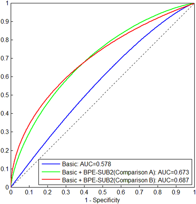
We investigated automated quantitative measures of background parenchymal enhancement (BPE) derived from an early versus delayed post-contrast sequence in breast dynamic contrast-enhanced magnetic resonance imaging (DCE-MRI) for association with breast cancer presence in a case-control study. DCE-MRIs were retrospectively analyzed for 51 cancer cases and 51 controls with biopsy-proven benign lesions, matched by age and year-of-MRI. BPE was quantified using fully-automated validated computer algorithms, separately from three sequential DCE-MRI post-contrast-subtracted sequences (SUB1, SUB2, and SUB3). The association of BPE computed from the three SUBs and other known factors with breast cancer were assessed in terms of odds ratio (OR) and area under the receiver operating characteristic curve (AUC). The OR of breast cancer for the percentage BPE measure (BPE%) quantified from SUB1 was 3.5 (95% Confidence Interval: 1.3, 9.8; p = 0.015) for 20% increments. Slightly lower and statistically significant ORs were also obtained for BPE quantified from SUB2 and SUB3. There was no significant difference (p > 0.2) in AUC for BPE quantified from the three post-contrast sequences and their combination. Our study showed that quantitative measures of BPE are associated with breast cancer presence and the association was similar across three breast DCE-MRI post-contrast sequences.
Conflict of interest statement
Wendie A. Berg is a consultant for data analysis and manuscript preparation for SuperSonic, Imagine. Dr. Berg is voluntary Chief Scientific Advisor to
Figures


Similar articles
-
Breast MRI contrast enhancement kinetics of normal parenchyma correlate with presence of breast cancer.Breast Cancer Res. 2016 Jul 22;18(1):76. doi: 10.1186/s13058-016-0734-0. Breast Cancer Res. 2016. PMID: 27449059 Free PMC article.
-
Ultrafast DCE-MRI for discriminating pregnancy-associated breast cancer lesions from lactation related background parenchymal enhancement.Eur Radiol. 2023 Nov;33(11):8122-8131. doi: 10.1007/s00330-023-09805-8. Epub 2023 Jun 6. Eur Radiol. 2023. PMID: 37278853
-
Quantitative assessment of background parenchymal enhancement in breast MRI predicts response to risk-reducing salpingo-oophorectomy: preliminary evaluation in a cohort of BRCA1/2 mutation carriers.Breast Cancer Res. 2015 May 19;17:67. doi: 10.1186/s13058-015-0577-0. Breast Cancer Res. 2015. PMID: 25986460 Free PMC article.
-
The Association of Background Parenchymal Enhancement at Breast MRI with Breast Cancer: A Systematic Review and Meta-Analysis.Radiology. 2019 Sep;292(3):552-561. doi: 10.1148/radiol.2019182441. Epub 2019 Jun 25. Radiology. 2019. PMID: 31237494
-
Ultrafast Dynamic Contrast-Enhanced MRI of the Breast: From Theory to Practice.J Magn Reson Imaging. 2024 Aug;60(2):401-416. doi: 10.1002/jmri.29082. Epub 2023 Dec 12. J Magn Reson Imaging. 2024. PMID: 38085134 Review.
Cited by
-
Breast cancer and background parenchymal enhancement at breast magnetic resonance imaging: a meta-analysis.BMC Med Imaging. 2021 Feb 19;21(1):32. doi: 10.1186/s12880-021-00566-8. BMC Med Imaging. 2021. PMID: 33607959 Free PMC article.
-
Quantitative background parenchymal enhancement to predict recurrence after neoadjuvant chemotherapy for breast cancer.Sci Rep. 2019 Dec 16;9(1):19185. doi: 10.1038/s41598-019-55820-5. Sci Rep. 2019. PMID: 31844135 Free PMC article.
-
Association of Breast Cancer Odds with Background Parenchymal Enhancement Quantified Using a Fully Automated Method at MRI: The IMAGINE Study.Radiology. 2023 Sep;308(3):e230367. doi: 10.1148/radiol.230367. Radiology. 2023. PMID: 37750771 Free PMC article.
-
Assessment of Quantitative Magnetic Resonance Imaging Background Parenchymal Enhancement Parameters to Improve Determination of Individual Breast Cancer Risk.J Comput Assist Tomogr. 2019 Jan/Feb;43(1):85-92. doi: 10.1097/RCT.0000000000000774. J Comput Assist Tomogr. 2019. PMID: 30052617 Free PMC article.
-
Background Parenchymal Enhancement on Breast MRI as a Prognostic Surrogate: Correlation With Breast Cancer Oncotype Dx Score.Front Oncol. 2021 Feb 4;10:595820. doi: 10.3389/fonc.2020.595820. eCollection 2020. Front Oncol. 2021. PMID: 33614481 Free PMC article.
References
-
- Saslow D, Boetes C, Burke W. American Cancer Society Breast Cancer Advisory Group: American Cancer Society guidelines for breast screening with MRI as an adjunct to mammography. CA: A Cancer Journal for Clinicians. 2007;57:75–89. - PubMed
Publication types
MeSH terms
Grants and funding
LinkOut - more resources
Full Text Sources
Other Literature Sources
Medical
