

Immediate Surgical Management of Traumatic Dislocation of the Eye Globe into the Maxillary Sinus: Report of a Rare Case and Literature Review
- PMID: 28523089
- PMCID: PMC5435495
- DOI: 10.1055/s-0036-1584393
Immediate Surgical Management of Traumatic Dislocation of the Eye Globe into the Maxillary Sinus: Report of a Rare Case and Literature Review
Abstract
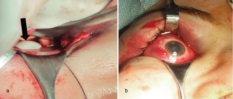
We report a case of complete dislocation of the globe into the maxillary sinus, with immediate repositioning of the globe. This report highlights the importance of early surgical repair of orbital fracture and globe repositioning to regain the maximum amount of ocular functions. A review of literature found 19 cases of globe dislocation into the maxillary sinus: One case was enucleated 2 months after misdiagnosis as traumatic enucleation, six cases were documented no vision or no light perception, three cases did not have reported vision (patients did not survive), and nine cases with postoperative vision. We recommend early surgical intervention to restore the cosmetic and visual function of the dislocated eye.
Keywords: eye; globe dislocation; optic nerve; orbital fracture.
Figures


References
-
- Ahmad F, Kirkpatrick W N, Lyne J, Urdang M, Garey L J, Waterhouse N. Strain gauge biomechanical evaluation of forces in orbital floor fractures. Br J Plast Surg. 2003;56(1):3–9. - PubMed
-
- Jellab B Baha A T Moutaouakil A et al. Management of a severe cranio-orbito-facial trauma with a dislocation of the globe into the maxillary sinus Bull Soc Belge Ophtalmol 2008309–310(309–310):37–41. - PubMed
-
- Morris W R, Osborn F D, Fleming J C. Traumatic evulsion of the globe. Ophthal Plast Reconstr Surg. 2002;18(4):261–267. - PubMed
Publication types
LinkOut - more resources
Full Text Sources
Other Literature Sources