Review
. 2017 Jul;13(7):420-433.
doi: 10.1038/nrneurol.2017.69. Epub 2017 May 19.

Modulators of microglial activation and polarization after intracerebral haemorrhage

Affiliations
Review

Modulators of microglial activation and polarization after intracerebral haemorrhage

Xi Lan et al. Nat Rev Neurol. 2017 Jul.

Abstract

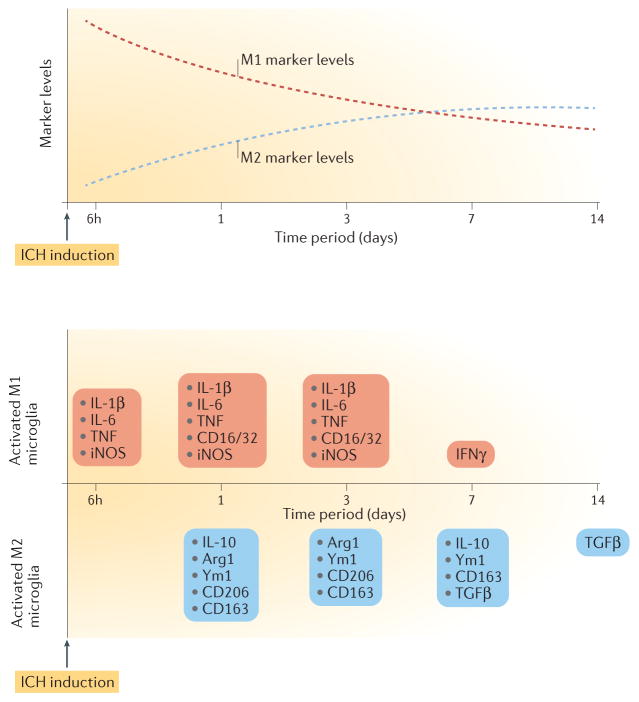
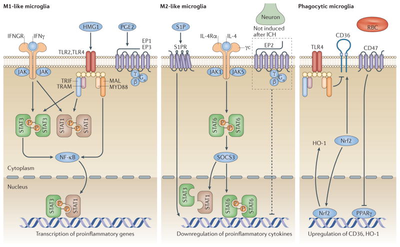
Intracerebral haemorrhage (ICH) is the most lethal subtype of stroke but currently lacks effective treatment. Microglia are among the first non-neuronal cells on the scene during the innate immune response to ICH. Microglia respond to acute brain injury by becoming activated and developing classic M1-like (proinflammatory) or alternative M2-like (anti-inflammatory) phenotypes. This polarization implies as yet unrecognized actions of microglia in ICH pathology and recovery, perhaps involving microglial production of proinflammatory or anti-inflammatory cytokines and chemokines. Furthermore, alternatively activated M2-like microglia might promote phagocytosis of red blood cells and tissue debris, a major contribution to haematoma clearance. Interactions between microglia and other cells modulate microglial activation and function, and are also important in ICH pathology. This Review summarizes key studies on modulators of microglial activation and polarization after ICH, including M1-like and M2-like microglial phenotype markers, transcription factors and key signalling pathways. Microglial phagocytosis, haematoma resolution, and the potential crosstalk between microglia and T lymphocytes, neurons, astrocytes, and oligodendrocytes in the ICH brain are described. Finally, the clinical and translational implications of microglial polarization in ICH are presented, including the evidence that therapeutic approaches aimed at modulating microglial function might mitigate ICH injury and improve brain repair.


Conflict of interest statement

Competing interests statement

The authors declare no competing interests.

Figures

Figure 1
Figure 1. Dynamic changes in microglial marker levels and profiles over time after intracerebral haemorrhage
Top panel, dashed red curve: microglial M1-like response exhibits a decreasing trend in the first 14 days. Top panel, dashed blue curve: microglial M2 like response exhibits an increasing trend in the first 14 days. Bottom panel: microglia exhibit an M1-like response as early as 6 h after intracerebral haemorrhage (ICH), as shown by upregulation of proinflammatory cytokines such as IL-1β, IL-6, tumour necrosis factor (TNF) and inducible nitric oxide synthase (iNOS). M2 markers, such as arginase-1 (Arg1), chitinase-like protein 3 (also known as Ym1), CD206, CD163 and IL-10, start to increase on day 1 after ICH. Transforming growth factor-β (TGFβ) is upregulated from day 7 until day 14 post-ICH. Although a mixed M1-like and M2-like microglial phenotype is evident during days 1 to 3, the balance of evidence supports an M1 to M2 phenotype switch in the first 7 days. IFNγ levels increase on day 7 after ICH, and levels of most proinflammatory cytokines return to baseline on day 14.
Figure 2
Figure 2. Modulators of microglial polarization and phagocytosis after intracerebral haemorrhage
Several transcription factors modulate microglial polarization towards an M1-like (left panel) or an M2-like (centre panel) phenotype. Activation of high-mobility group protein 1 (HMG1) and Toll-like receptor (TLR)2 or TLR4 promotes microglial M1-like responses. Signal transducer and activator of transcription (STAT)1 is activated by TLRs and IFNγ, promoting microglial polarization to an M1 phenotype. After intracerebral haemorrhage (ICH), TLRs modulate STAT3 phosphorylation, which increases M1-like polarization. G-protein-coupled E prostanoid (EP) receptors EP1 and EP3 are mainly expressed on microglia and drive neuronal toxicity mediated by M1 upregulation after ICH. STAT6 accumulates in response to IL-4 and is responsible for the transcription of M2-related genes. Sphingosine-1-phosphate (S1P) receptor signalling contributes to the downregulation of proinflammatory cytokines and enhances M2-like responses after ICH. Although EP2 receptors are not expressed in microglia after ICH, EP2 deletion results in an increased microglial proinflammatory response. Other modulators regulate microglial phagocytosis (right panel). Activation of the transcription factor nuclear factor erythroid 2 related factor 2 (Nrf2) is associated with microglial phagocytosis after ICH and increases CD36 and haem oxygenase-1 (HO-1) expression. The CD36 gene is a target of peroxisome proliferator-activated receptor-γ (PPARγ), which belongs to the nuclear receptor family. CD47 expression in red blood cells (RBC) can decrease their phagocytosis by microglia after ICH. Although CD36 expression is increased in the ICH brain, TLR4 activation might negatively regulate its expression and delay haematoma clearance. IFGNR, IFNγ receptor; NF-κB, nuclear factor-κB; MAL, Toll/IL-1 receptor domain-containing adaptor protein (also known as MYD88 adapter-like protein); MYD88, myeloid differentiation primary response protein MYD88; SOCS3, suppressor of cytokine signalling 3; TRAM, TIR domain-containing adaptor molecule 2 (also known as TRIF-related adaptor molecule); TRIF, TIR domain-containing adaptor molecule 1.

References

    1. Feigin VL, Lawes CM, Bennett DA, Barker-Collo SL, Parag V. Worldwide stroke incidence and early case fatality reported in 56 population-based studies: a systematic review. Lancet Neurol. 2009;8:355–369. - PubMed
    1. Steiner T, et al. European Stroke Organisation (ESO) guidelines for the management of spontaneous intracerebral hemorrhage. Int J Stroke. 2014;9:840–855. - PubMed
    1. Kim JY, Bae HJ. Spontaneous intracerebral hemorrhage: management. J Stroke. 2017;19:28–39. - PMC - PubMed
    1. Thabet AM, Kottapally M, Hemphill JC., III Management of intracerebral hemorrhage. Handb Clin Neurol. 2017;140:177–194. - PubMed
    1. Hemphill JC, III , et al. Guidelines for the management of spontaneous intracerebral hemorrhage: a guideline for healthcare professionals from the American Heart Association/American Stroke Association. Stroke. 2015;46:2032–2060. - PubMed