. 2017 Nov;21(11):2796-2808.
doi: 10.1111/jcmm.13194. Epub 2017 May 19.

Polydatin reduces Staphylococcus aureus lipoteichoic acid-induced injury by attenuating reactive oxygen species generation and TLR2-NFκB signalling

Affiliations

Polydatin reduces Staphylococcus aureus lipoteichoic acid-induced injury by attenuating reactive oxygen species generation and TLR2-NFκB signalling

Gan Zhao et al. J Cell Mol Med. 2017 Nov.

Abstract

Staphylococcus aureus (S. aureus) causes severe inflammation in various infectious diseases, leading to high mortality. The clinical application of antibiotics has gained a significant curative effect. However, it has led to the emergence of various resistant bacteria. Therefore, in this study, we investigated the protective effect of polydatin (PD), a traditional Chinese medicine extract, on S. aureus lipoteichoic acid (LTA)-induced injury in vitro and in vivo. First, a significant improvement in the pathological conditions of PD in vivo was observed, suggesting that PD had a certain protective effect on LTA-induced injury in a mouse model. To further explore the underlying mechanisms of this protective effect of PD, LTA-induced murine macrophages were used in this study. The results have shown that PD could reduce the NF-κB p65, and IκBα phosphorylation levels increased by LTA, resulting in a decrease in the transcription of pro-inflammatory factors, such as TNF-α, IL-1β and IL-6. However, LTA can not only activate NF-κB through the recognition of TLR2 but also increase the level of intracellular reactive oxygen species (ROS), thereby activating NF-κB signalling. We also detected high levels of ROS that activate caspases 9 and 3 to induce apoptosis. In addition, using a specific NF-κB inhibitor that could attenuate apoptosis, namely NF-κB p65, acted as a pro-apoptotic transcription factor in LTA-induced murine macrophages. However, PD could inhibit the generation of ROS and NF-κB p65 activation, suggesting that PD suppressed LTA-induced injury by attenuating ROS generation and TLR2-NFκB signalling.

Keywords: ROS; NF-κB; apoptosis; inflammation.


Figures

Figure 1
Figure 1
(A) Chemical structure of polydatin. (B) Effect of polydatin on cell viability. Cells were treated with the indicated concentration of polydatin (0, 12.5, 25, 50, 100 μg/ml) for 24 hrs, and cell viability was detected by CCK‐8 kits.
Figure 2
Figure 2
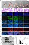
(A) Histological examination of the protective effect of polydatin on LTA‐induced uterine injury in mice (n = 6). From top to bottom: uterine morphology observation, scale bar: 1 cm; H&E staining of uterine tissue; phosphorylated NF‐κB p65 immunofluorescence staining (Green) of uterine tissue; TUNEL staining of uterine tissue. Cell nuclei (Blue), TUNEL‐positive cells (Red). Scale bar: 200 μm. The red, white and blue arrows indicate the tissue lesion, the translocation of p65 and the apoptotic region, respectively. (B) The protein levels of phosphorylated NF‐κB p65 (p‐p65), phosphorylated IκBα (p‐IκBα) and cleaved caspases 9 and 3 were determined by Western blotting. β‐Actin was used as an internal control. (C) The Western blotting data were represented the means ± S.E.M. of three independent experiments. CG is the control group, LTA is the LTA group, and 25, 50 and 100 are the polydatin‐treatment groups representing 25 mg/kg, 50 mg/kg and 100 mg/kg per animal, respectively. # P < 0.05, ## P < 0.01 versus the CG group. *P < 0.05, **P < 0.01 versus the LTA group.
Figure 3
Figure 3
Effect of polydatin on apoptosis induced by LTA. (A) Representative dot plots of staining with Annexin V and PI. Cells were treated as described previously. (B) Numbers in the quadrants are the percentages of each population. The data are represented as the means ± S.E.M. of three independent experiments. (C) The protein levels of cleaved caspases 9 and 3 were determined by Western blotting. β‐Actin was used as an internal control. (D) The Western blotting data were represented as the means ± S.E.M. of three independent experiments. CG is the control group, LTA is the LTA group, and 12.5, 25 and 50 are the polydatin‐treatment groups representing 12.5 μg/ml, 25 μg/ml and 50 μg/ml per cell plate, respectively. # P < 0.05, ## P < 0.01 versus the CG group. *P < 0.05, **P < 0.01 versus the LTA group.
Figure 4
Figure 4
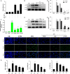
Fluorescence microscopy of ROS production by DCFHDA (green) after stimulation or treatment (ROS). Inhibition of LTA‐induced cell apoptosis by polydatin was examined by the TUNEL assay (TUNEL). Blue spots represent cell nuclei, and red spots represent TUNEL‐positive cells. CG is the control group, LTA is the LTA group, and 12.5, 25 and 50 are the polydatin‐treatment groups representing 12.5 μg/ml, 25 μg/ml and 50 μg/ml per cell plate, respectively. NAC is the NAC (500 μM)‐treatment group. The integrated option density (IOD) of DAPI was used as an internal control. All of the data represent the mean ± S.E.M. of three independent experiments. # P < 0.05, ## P < 0.01 versus the CG group. *P < 0.05, **P < 0.01 versus the positive LTA group.
Figure 5
Figure 5
(A) The interfering efficiency of TLR2 siRNA and effect of LTA or H2O2 on TLR2 expression were measured by RTPCR. (B) The protein levels of p‐p65 and p‐IκBα were stimulated with LTA after the knockdown of TLR2 by siRNA or PD pre‐treatment. (C) The protein levels of p‐p65 and p‐IκBα were stimulated with H2O2 after the knockdown of TLR2 by siRNA or PD or NAC pre‐treatment. β‐Actin was used as an internal control. (D, E) The Western blotting data were represented as the means ± S.E.M. of three independent experiments. (F) Translocation of the p65 subunit from the cytoplasm into the nucleus was evaluated by immunofluorescence. Blue spots represent cell nuclei, and green spots represent p‐p65 staining. The integrated option density (IOD) of DAPI was used as an internal control. (G) The effect of polydatin on the mRNA levels of IL‐1β, IL‐6 and TNF‐α induced by LTA was determined by qPCR in RAW 264.7 cells. GAPDH was used as a control. All of the data were represented as the means ± S.E.M. of three independent experiments. CG: Control group, LTA: LTA group, NAC: NAC‐treatment group, H2O2: H2O2 group, si‐TLR2: TLR2 siRNA, si‐NC: TLR2 siRNA negative control; PD(50): cells treated with polydatin with a concentration of 25 μg/ml, and 12.5, 25 and 50 represent the polydatin‐treatment groups representing 12.5 μg/ml, 25 μg/ml and 50 μg/ml per cell plate, respectively. Data represent the mean ± S.E.M. of three independent experiments. # P < 0.05, ## P < 0.01 versus the CG group. *P < 0.05, **P < 0.01 versus the positive group (LTA or H2O2).
Figure 6
Figure 6
(A) The protein levels of p‐p65 stimulated with LTA after blockade by BAY‐11‐7082 with the indicated concentration. β‐Actin was used as an internal control. (C) The protein levels of cleaved caspases 9 and 3 stimulated with LTA after blockade by BAY‐11‐7082 with the indicated concentrations. β‐Actin was used as an internal control. (B, D) The Western blotting data were represented as the means ± S.E.M. of three independent experiments. CG is the control group, LTA is the LTA group, and 0, 5, 10, 20 are the BAY‐11‐7082‐treatment groups representing the concentrations of 0, 5, 10 and 20 μM per cell plate, respectively. # P < 0.05, ## P < 0.01 versus the CG group. *P < 0.05, **P < 0.01 versus the LTA group.
Figure 7
Figure 7
Schematic diagram of a signalling pathway related to anti‐apoptotic or anti‐inflammatory effects of polydatin on LTA‐induced injury. LTA can induce NFκB activation in a TLR2‐dependent or TLR2‐independent manner, leading to the release of downstream pro‐inflammatory cytokines. Moreover, LTA can increase the level of intracellular ROS, which induce apoptosis via activating caspases 9 and 3. In addition, NFκB acts as a pro‐apoptotic regulator involved in apoptosis signalling. However, the treatment of PD can suppress LTA‐induced injury by attenuating ROS generation and TLR2‐NFκB activation.

References

    1. Kriebs JM. Staphylococcus Infections in Pregnancy: maternal and Neonatal Risks. J Perinat Neonatal Nurs. 2016; 30: 115–23. - PubMed
    1. Johnston JN, Kaplan SL, Mason EO, et al Characterization of Staphylococcus aureus infections in children with down syndrome. J Infect Chemother. 2015; 21: 790–4. - PubMed
    1. Zhao G, Wu H, Jiang K, et al IFN‐τ inhibits S. aureus‐induced inflammation by suppressing the activation of NF‐κB and MAPKs in RAW 264.7 cells and mice with pneumonia. Int Immunopharmacol. 2016; 35: 332–40. - PubMed
    1. Seixas R, Varanda D, Bexiga R, et al Biofilm‐formation by Staphylococcus aureus and Staphylococcus epidermidis isolates from subclinical mastitis in conditions mimicking the udder environment. Pol J Vet Sci. 2015; 18: 787–92. - PubMed
    1. Crum‐Cianflone NF, Wang X, Weintrob A, et al Specific behaviors predict Staphylococcus aureus colonization and skin and soft tissue infections among human immunodeficiency virus‐infected persons. Open Forum Infect Dis. 2015; 2: ofv034. - PMC - PubMed

MeSH terms

Associated data