Review
. 2017 Aug;52(4):425-460.
doi: 10.1080/10409238.2017.1325829. Epub 2017 May 19.

Ubiquitin enzymes in the regulation of immune responses

Affiliations
Review

Ubiquitin enzymes in the regulation of immune responses

Petra Ebner et al. Crit Rev Biochem Mol Biol. 2017 Aug.

Abstract

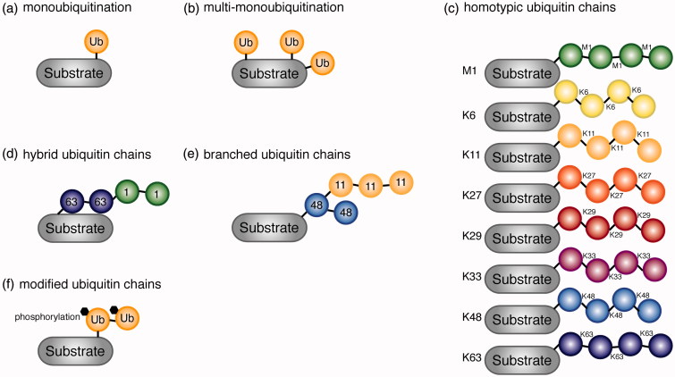
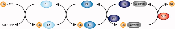
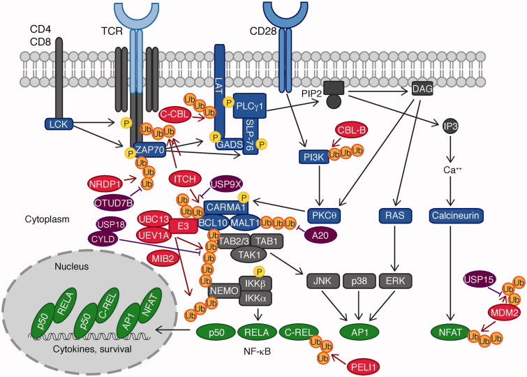
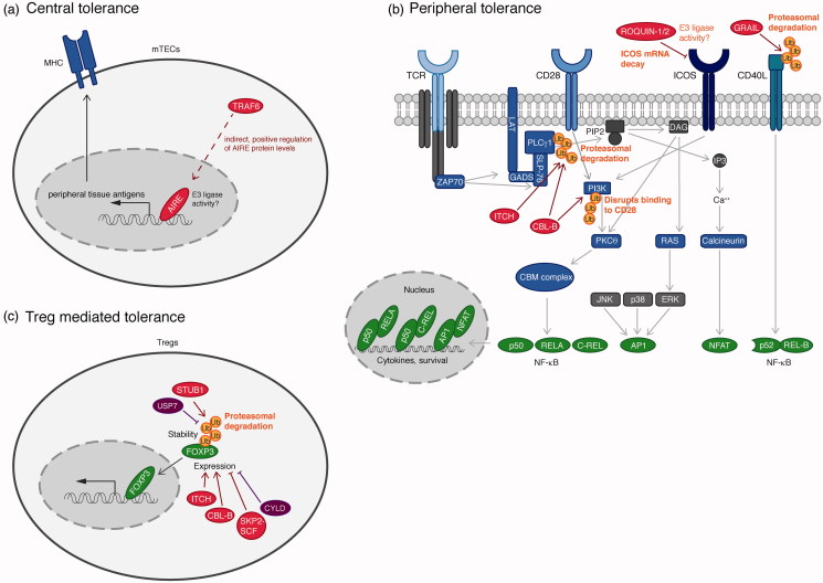
Ubiquitination plays a central role in the regulation of various biological functions including immune responses. Ubiquitination is induced by a cascade of enzymatic reactions by E1 ubiquitin activating enzyme, E2 ubiquitin conjugating enzyme, and E3 ubiquitin ligase, and reversed by deubiquitinases. Depending on the enzymes, specific linkage types of ubiquitin chains are generated or hydrolyzed. Because different linkage types of ubiquitin chains control the fate of the substrate, understanding the regulatory mechanisms of ubiquitin enzymes is central. In this review, we highlight the most recent knowledge of ubiquitination in the immune signaling cascades including the T cell and B cell signaling cascades as well as the TNF signaling cascade regulated by various ubiquitin enzymes. Furthermore, we highlight the TRIM ubiquitin ligase family as one of the examples of critical E3 ubiquitin ligases in the regulation of immune responses.

Keywords: B cell signaling; E3 ligase; T cell signaling; TNF signaling; TRIM; Ubiquitin; deubiquitinase.


Figures

Figure 1.
Figure 1.
Various types of ubiquitin signals are generated based on the linkage type. (a) Attachment of one ubiquitin molecule to a substrate, monoubiquitination. (b) Monoubiquitination on several lysine residues on the same substrate, multi-monoubiquitination. (c) Homotypic ubiquitin chains linked via intrinsic Met 1 and Lys residues (M1, K6, K11, K27, K29, K33, K48, and K63). (d) Hybrid ubiquitin chains consisting of multiple linkage types of chains. K63-M1 hybrid chain is shown. (e) Branched ubiquitin chain consisting of K48 and K11 linkages. (f) Modified (phosphorylated) ubiquitin moiety forming ubiquitin chains on the substrate (see color version of this figure at www.tandfonline.com/ibmg).
Figure 2.
Figure 2.
Ubiquitination is a three-step enzymatic process. A ubiquitin-activating enzyme (E1), a ubiquitin-conjugating enzyme (E2), and a ubiquitin ligase (E3) act together to form a covalent bond between ubiquitin and its substrate protein. The E1 enzyme uses ATP to form a thioester bond between its active site cysteine and the C-terminal glycine of ubiquitin. The ubiquitin is then transferred on to the cysteine in the active site of the E2 enzyme, which cooperates with three classes of E3 enzymes to conjugate ubiquitin on the substrate. Deubiquitinases (DUBs) reverse the ubiquitination reaction and hydrolyze ubiquitin from the substrate (see color version of this figure at www.tandfonline.com/ibmg).
Figure 3.
Figure 3.
Ubiquitin enzymes in the TCR signaling pathway. Antigen binding to the TCR leads to the recruitment of the tyrosine kinase LCK, which phosphorylates the TCR signaling chain CD3ζ, which recruits ZAP70. Subsequent phosphorylation of LAT and SLP-76 by ZAP70 triggers activation of PLCγ1, cleaving PIP2 to IP3, and DAG. IP3 and DAG activate NF-κB, AP-1, and NFAT via the protein kinase PKCθ, RAS, and calcium, respectively. NF-κB and AP-1 activation is mediated by the CBM complex, consisting of CARMA1, BCL10, and MALT1, which cooperates with an E3 and the Lys 63-specific E2 dimer UBC13/UEV1A. Ubiquitination of BCL10 leads to the recruitment of the TAB2/TAB1/TAK1 complex and the IKK complex for NF-κB and JNK activation. E3 ligases are indicated in red, DUBs in purple, and transcription factors in green (see color version of this figure at www.tandfonline.com/ibmg).
Figure 4.
Figure 4.
Ubiquitin enzymes in the regulation of T cell-mediated autoimmunity. (a) The transcription factor AIRE, crucial for central tolerance, controls expression of peripheral tissue antigens, which are required for the negative selection process. An involvement of the E3 ligase activity of AIRE has not been clarified. The E3 ligase TRAF6 indirectly controls AIRE expression via the canonical NF-κB pathway. (b) Peripheral tolerance ensures that self-reactive T cells enter an inactive state (anergy). The E3 ligases ITCH and CBL-B ubiquitinate PLCγ1 and the regulatory subunit of PI3K, p85. Lys 48-linked ubiquitination of PLCγ1 induces proteasomal degradation, whereas ubiquitination of PI3K blocks recruitment to CD28, both of which causes termination of TCR signaling. The E3 ligase GRAIL induces ubiquitination of the co-stimulatory molecule CD40 ligand, which induces its proteasomal degradation. The E3 ligases ROQUIN-1 and ROQUIN-2 control levels of ICOS by inducing ICOS mRNA decay. (c) Treg generation is controlled by the transcription factor FOXP3. The ubiquitin E3 ligase STUB1 directly ubiquitinates FOXP3 with Lys 48-linked ubiquitin chains, leading to proteasomal degradation, which can be counteracted by the DUB USP7. CBL-B and ITCH indirectly promote Treg generation by positively regulating FOXP3 expression. Contrarily, the SKP2-SCF complex as well as the DUB CYLD induce loss of FOXP3 expression via indirect effects. E3 ligases are indicated in red, DUBs in purple, and transcription factors in green (see color version of this figure at www.tandfonline.com/ibmg).
Figure 5.
Figure 5.
Ubiquitination in B cell activation. In B cells, the non-canonical NF-κB pathway mediated by two TNF receptor superfamily members, CD40 and BAFF receptor (BAFFR) is regulated by ubiquitination. Upon activation of CD40 or BAFFR, TRAF2/6 promotes activation of cIAP1-cIAP2, which in turn mediates Lys 48-linked ubiquitination and degradation of TRAF3. Degradation of TRAF3 results in stabilization of NIK. Active NIK phosphorylates and activates IKKα, which in turn phosphorylates the NF-κB subunit p100 (a precursor of p52). Phosphorylation-induced ubiquitination of p100 induces processing by the proteasome. This releases the mature p52 subunit, which associates with REL-B to promote expression of genes required for B cell survival, maturation and activation. Degradation of TRAF3 results in cytosolic translocation of a signaling complex containing the MAPK kinase kinase MEKK1. MEKK1 then activates JNK. An additional layer of regulation is provided by the E3 ligase ACT1 as well as the DUB OTUD7B. E3 ligases are indicated in red, DUBs in purple, and transcription factors in green (see color version of this figure at www.tandfonline.com/ibmg).
Figure 6.
Figure 6.
Ubiquitin chains and ubiquitin enzymes in the TNF-induced NF-κB and apoptosis pathways. Different linkage types of ubiquitin chains, Met 1-, Lys 11-, Lys 48-, and Lys 63-linked ubiquitin chains play a critical role in the TNF-induced canonical NF-κB and the TNFR complex II-dependent apoptosis pathways. Ubiquitin chains are generated by the E3 ligases, cIAP, LUBAC complex, and SCF-βTrCP. These ubiquitin chains are hydrolyzed by two DUBs, OTULIN and CYLD. A20 is a hybrid of E3 ligase and DUB. Ubiquitination of the substrates, including cIAPs, RIPK1, NEMO, and IκB-α, impacts on the downstream signaling pathways. The TNFR complex II-mediated apoptosis pathway includes RIPK1, TRADD, FADD, and Caspase 8. Activation of Caspase 8 leads to Caspase 3-dependent cleavage of PARP and apoptosis. The LUBAC complex (HOIP, SHARPIN, and HOIL-1L) and the CYLD–SPATA2 complex regulate the TNFR complex II-induced apoptosis pathway. E3 ligases are indicated in red, DUBs in purple, and transcription factors in green (see color version of this figure at www.tandfonline.com/ibmg).
Figure 7.
Figure 7.
TRIMs in the regulation of the immune response. (a) TRIM5α restricts retroviral infection. The TRIM5α SPRY domain interacts with the retroviral capsid lattice, which has two distinct consequences. First, TRIM5α recruits the autophagy machinery for degradation of the capsid. Second, TRIM5α in conjunction with UEV1A/UBC13 generates unanchored Lys 63-linked ubiquitin chains, which mediates TAK1 transactivation and subsequent NF-κB activation. (b) TRIM25 positively controls IFNβ production. By generating Lys 63-linked ubiquitin chains, TRIM25 releases RIG-I from its repressed conformation, which results in RIG-I tetramerization and subsequent IFNβ production. (c) TRIM6 controls type I interferon production and signaling, by generating Lys 48-linked, unanchored ubiquitin chains, which activate IKKɛ. Nipah virus antagonizes interferon signaling by targeting TRIM6 for degradation. (d) TRIM65 activates the cytoplasmic dsRNA sensor MDA5 by conjugation of Lys 63-linked ubiquitin chains on MDA5 Lys 743. TRIM65 is critical for MDA5 oligomerization and activation. Similar to TRIM56, Salmonella SopA can also interact with TRIM65 and mediate its degradation. However, unlike TRIM56, SopA does not interfere with TRIM65 E3 activity. (e) TRIM56 controls the STING-dependent cytosolic dsDNA response pathway by ubiquitinating STING with Lys 63-linked ubiquitin chains on Lys 150. Ubiquitination allows for STING dimerization, which is crucial for its activation. Salmonella SopA has been shown to bind and ubiquitinate TRIM56, thereby inhibiting it through preventing E3 ligase activity and degradation, respectively (see color version of this figure at www.tandfonline.com/ibmg).

Similar articles

Cited by

References

    1. Abramson J. and Goldfarb Y., 2016. AIRE: from promiscuous molecular partnerships to promiscuous gene expression. European journal of immunology, 46, 22–33. - PubMed
    1. Aggarwal B.B., 2003. Signalling pathways of the TNF superfamily: a double-edged sword. Nature reviews immunology, 3, 745–756. - PubMed
    1. Ahn J., Ruiz P., and Barber G.N., 2014. Intrinsic self-DNA triggers inflammatory disease dependent on STING. Journal of immunology, 193, 4634–442. - PMC - PubMed
    1. Aireakiyama T., et al, 2008. The tumor necrosis factor family receptors RANK and CD40 cooperatively establish the thymic medullary microenvironment and self-tolerance. Immunity, 29, 423–437. - PubMed
    1. Akiyama T., et al, 2005. Dependence of self-tolerance on TRAF6-directed development of thymic stroma. Science, 308, 248–251. - PubMed

LinkOut - more resources