

Micro-CT analysis of the rodent jaw bone micro-architecture: A systematic review
- PMID: 28525530
- PMCID: PMC5365162
- DOI: 10.1016/j.bonr.2014.10.005
Micro-CT analysis of the rodent jaw bone micro-architecture: A systematic review
Abstract
Introduction: Knowledge about macro- and micro-structural characteristics may improve in vivo estimation of the quality and quantity of regenerated bone tissue. For this reason, micro-CT imaging has been applied to evaluate alveolar bone remodelling, alterations of periodontal ligament thickness and cortical and trabecular bone changes in rodent jaw bones. In this paper, we provide a systematic review on the available micro-CT literature on jaw bone micro-architecture.
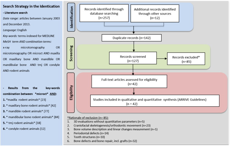
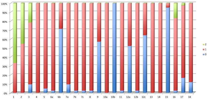
Methodology: A detailed search through the PubMed database was performed. Articles published up to December 2013 and related to maxilla, mandible and condyle with quantitatively analysed bone micro-architectural parameters were considered eligible for inclusion. Two reviewers assessed the search results according to inclusion criteria designed to identify animal studies quantifying the bone micro-architecture of the jaw rodent bones in physiological or drug-induced disease status, or in response to interventions such as mechanical loading, hormonal treatment and other metabolic alterations. Finally, the reporting quality of the included publications was evaluated using the tailored ARRIVE guidelines outlined by Vignoletti and Abrahamsson (2012).
Results: Database search, additional manual searching and assessment of the inclusion and exclusion criteria retrieved 127 potentially relevant articles. Eventually, 14 maxilla, 20 mandible and 12 condyle articles with focus on bone healing were retained, and were analysed together with 3 methodological papers. Each study was described systematically in terms of subject, experimental intervention, follow-up period, selected region of interest used in the micro-CT analysis, parameters quantified, micro-CT scanner device and software. The evidence level evaluated by the ARRIVE guidelines showed high mean scores (between 18 and 25; range: 0-25), indicating that most of the selected studies are well-reported. The major obstacles identified were related to sample size calculation, absence of adverse event descriptions, randomization or blinding procedures.
Conclusions: The evaluated studies are highly heterogeneous in terms of research topic and the different regions of interest. These results illustrate the need for a standardized methodology in micro-CT analysis. While the analysed studies do well according to the ARRIVE guidelines, the micro-CT procedure is often insufficiently described. Therefore we recommend to extend the ARRIVE guidelines for micro-CT studies.
Keywords: Bone micro-architecture; Condyle; Mandible; Maxilla; Micro-CT; Rodent jaws.
Figures
References
-
- Muller R. Hierarchical microimaging of bone structure and function. Nat. Rev. Rheumatol. 2009;5(7):373–381. - PubMed
-
- Bouxsein M.L. Guidelines for assessment of bone microstructure in rodents using micro-computed tomography. J. Bone Miner. Res. 2010;25(7):1468–1486. - PubMed
-
- Dai Q.G., Zhang P. Ovariectomy induces osteoporosis in the maxillary alveolar bone: an in vivo micro-CT and histomorphometric analysis in rats. Oral Dis. 2014;20(5):514–520. - PubMed
-
- Kallai I. Microcomputed tomography-based structural analysis of various bone tissue regeneration models. Nat. Protoc. 2011;6(1):105–110. - PubMed
Publication types
LinkOut - more resources
Full Text Sources
Other Literature Sources
