. 2017 May 18;66(4):503-516.e5.
doi: 10.1016/j.molcel.2017.04.028.

Ubiquitin Modification by the E3 Ligase/ADP-Ribosyltransferase Dtx3L/Parp9

Affiliations

Ubiquitin Modification by the E3 Ligase/ADP-Ribosyltransferase Dtx3L/Parp9

Chun-Song Yang et al. Mol Cell. .

Abstract

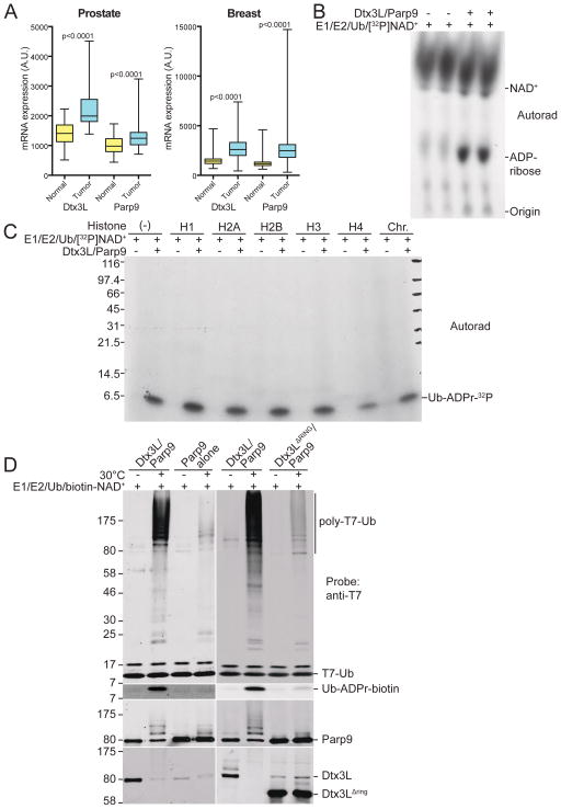
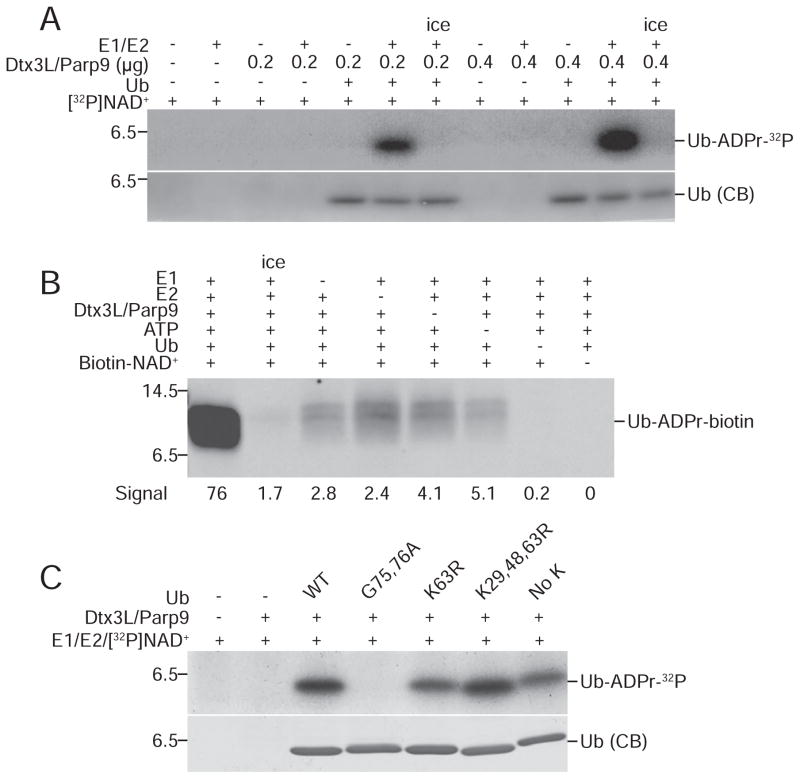
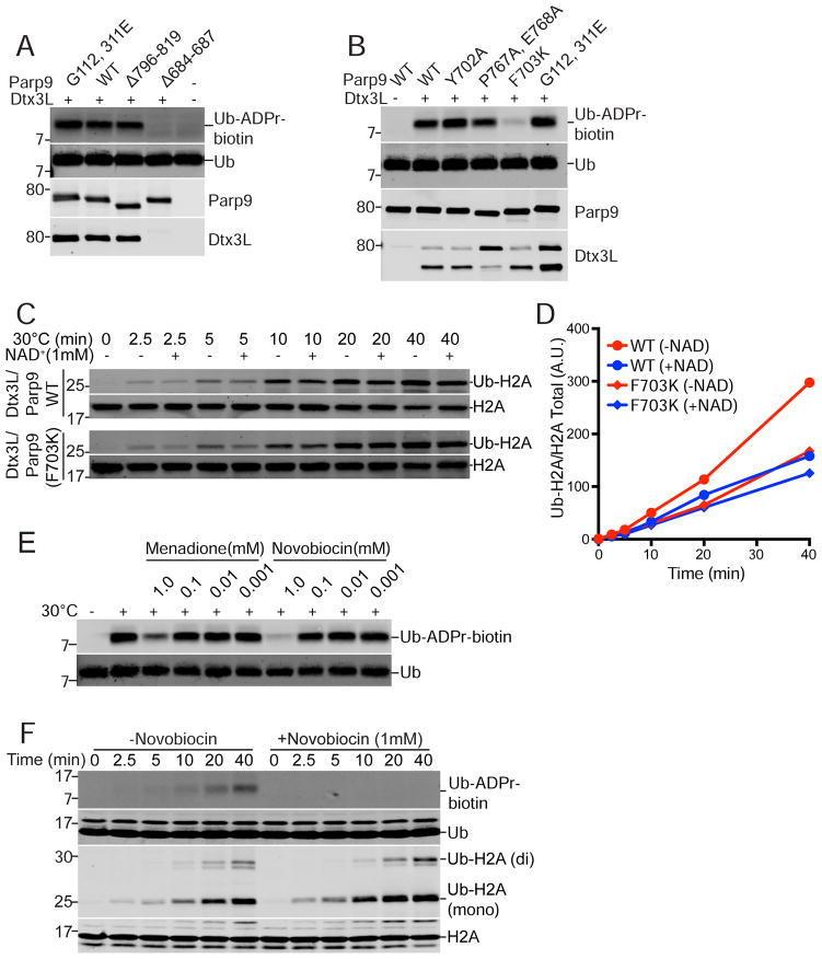
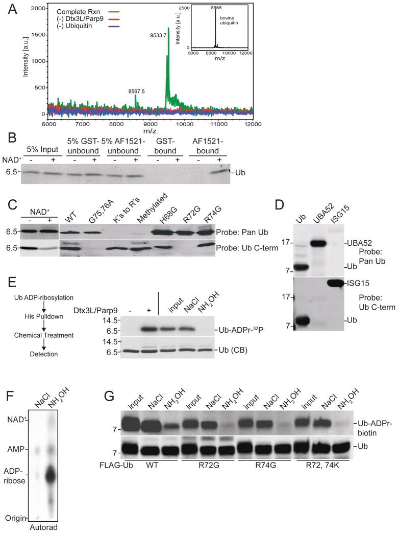
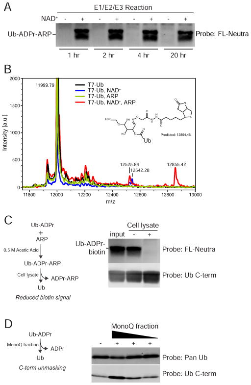
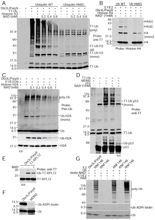
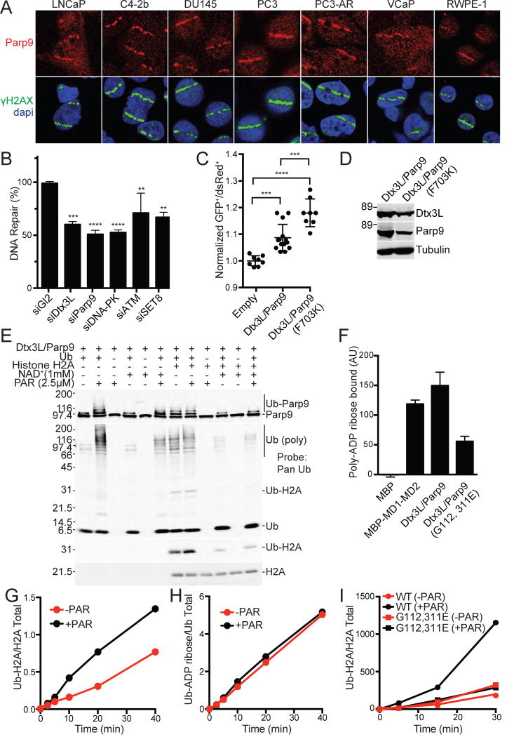
ADP-ribosylation of proteins is emerging as an important regulatory mechanism. Depending on the family member, ADP-ribosyltransferases either conjugate a single ADP-ribose to a target or generate ADP-ribose chains. Here we characterize Parp9, a mono-ADP-ribosyltransferase reported to be enzymatically inactive. Parp9 undergoes heterodimerization with Dtx3L, a histone E3 ligase involved in DNA damage repair. We show that the Dtx3L/Parp9 heterodimer mediates NAD+-dependent mono-ADP-ribosylation of ubiquitin, exclusively in the context of ubiquitin processing by E1 and E2 enzymes. Dtx3L/Parp9 ADP-ribosylates the carboxyl group of Ub Gly76. Because Gly76 is normally used for Ub conjugation to substrates, ADP-ribosylation of the Ub carboxyl terminus precludes ubiquitylation. Parp9 ADP-ribosylation activity therefore restrains the E3 function of Dtx3L. Mutation of the NAD+ binding site in Parp9 increases the DNA repair activity of the heterodimer. Moreover, poly(ADP-ribose) binding to the Parp9 macrodomains increases E3 activity. Dtx3L heterodimerization with Parp9 enables NAD+ and poly(ADP-ribose) regulation of E3 activity.


Figures

Figure 1
Figure 1. Expression and activity of the Dtx3L/Parp9 heterodimer
(A) Box and whisker plots showing expression of Dtx3L and Parp9 in cancer (blue) compared to adjacent normal tissue (yellow) for prostate and breast cancer, and nine additional cancers (Figure S1). RNA-Seq by Expectation-Maximization (RSEM) values from TCGA were used to plot normalized expression, whiskers represent the minimum and maximum data values, and significance was calculated using a paired t-test. (B) Recombinant Dtx3L/Parp9 cleaves NAD+. Ubiquitylation reactions containing 32P-NAD+ were analyzed by TLC and autoradiography, with unlabeled ADP-ribose as a migration standard. (C) Dtx3L/Parp9 ADP-ribosylates Ub. Ubiquitylation reactions with purified Dtx3L/Parp9, histones, and chromatin in the presence of 32P-NAD+. The reactions were subjected to SDS-PAGE, Coomassie Blue (CB) staining (Figure S3D), and autoradiography. (D) Ubiquitylation reactions with different combinations of human Dtx3L and Parp9 purified from insect cells. Biotin-labeled NAD+ was added to monitor ADP-ribosylation of Ub (FL-Neutra detection).
Figure 2
Figure 2. ADP-ribosylation of Ub by Dtx3L/Parp9 requires E1 and E2 processing
(A) ADP ribosylation reactions were performed using 32P-NAD+ and analyzed by SDS-PAGE and autoradiography with the protein combinations indicated. (B) ADP-ribosylation reactions were performed using biotin-NAD+ with drop-out of individual components as indicated. ADP-ribosylation of Ub was measured by probing with FL-Neutra. (C) ADP-ribosylation reactions were performed using 32P-NAD+ and Ub mutants bearing amino acid substitutions that affect processing (G75, 76A) and conjugation (K63R; K29, 48, 63R; No K).
Figure 3
Figure 3. Ub ADP-ribosylation mediated by the Parp9 catalytic domain
(A) Parp9 mutations engineered in the macrodomains (G112, 311E) and catalytic domain (Δ796-819; Δ684-687) expressed in E. coli and tested for Ub ADP-ribosylation (ADPr-biotin) and heterodimerization with Dtx3L. The locations of the mutations are depicted within a structural model of Parp9 (Figure S4). (B) Parp9 mutations engineered in the catalytic domain (Y702A; P767A, E768A; F703K) expressed in mammalian cells and tested for Ub ADP-ribosylation (ADPr-biotin) and heterodimerization with Dtx3L. (C, D) E3 activity of WT Parp9 and F703K Parp9 using recombinant heterodimers with Dtx3L. (E) Effect of non-selective Parp inhibitors Menadione and Novobiocin on Ub ADP-ribosylation. (F) Time course of Ub-ADP-ribosylation and H2A ubiquitylation in the presence of Novobiocin.
Figure 4
Figure 4. Dtx3L/Parp9 ADP-ribosylates the C-terminus of Ub
(A) Spectra from MALDI-TOF analysis of Ub. ADP-ribosylation reactions were performed with biotin-NAD+ with drop-out of Dtx3L/Parp9 and purified bovine Ub (inset shows unmodified Ub). ADPr-biotin labeled reaction product was isolated on streptavidin beads and analyzed by MS. (B) Recovery of ADP-ribosylated Ub on recombinant Af1521 macrodomain. Ub ADP-ribosylation reactions performed in the absence and presence of unlabeled NAD+ were dialyzed and then combined with GST- and GST-Af1521 beads. The input, unbound, and bound fractions were analyzed by immunoblotting with a pan-Ub Ab. (C) ADP-ribosylation of Ub reduces C-term antibody binding. ADP-ribosylation reactions were performed in the absence and presence of unlabeled NAD+ and Ub immunoblotting performed with Pan-Ub and Ub C-term Abs (left panel). Epitope mapping of Pan-Ub and Ub C-term Abs using WT and mutant Ub proteins (right panel). (D) Mapping the C-term Ab epitope with Ub proteins. Recombinant Ub, Uba52, or ISG15 (mature form) was separated via SDS-PAGE and probed with Pan-Ub antibody or Ub C-term antibody. Alignments of the C-terminal domains of the Ub proteins are provided (Figure S6C). (E) Chemical sensitivity of ADP-ribose conjugated to Ub. ADP-ribosylation reactions containing His-Ub and 32P-NAD+ were recovered on cobalt beads, treated as indicated, and analyzed by SDS-PAGE and autoradiography. (F) Release of 32P-ADPr from Ub by NH2OH treatment analyzed by TLC. (G) WT and Ub mutants were ADP-ribosylated using biotin-NAD+ and ADPr release determined by probing with FL-Neutra. ADPr release from Ub is CHES-resistant (Figure S6E).
Figure 5
Figure 5. ADP-ribosylated Ub is reactive with N-(aminooxyacetyl)-N'-(D-Biotinoyl) hydrazine (ARP)
(A) ARP modification of Ub requires initial ADP-ribosylation. The E1/E2/E3 reactions were performed in the absence and presence of NAD+. In a second step, ARP reactions were performed at room temperature for the times indicated, followed by FL-Neutra detection of the biotin moiety in ARP. (B) Spectra of reactions containing combinations of Ub, NAD+, and ARP. The 12855.42 Da mass is unique to T7-Ub-ADPr labeled with ARP. A putative structure consistent with the mass is shown, along with a potential mechanism (Figure S6F). (C) ADP-ribosylation of Ub is reversible. T7-Ub-ADPr-ARP was generated, incubated with cell buffer or cell lysate, and subsequently assayed for the loss of ADPr-ARP by streptavidin detection of biotin. (D) Removal of ADPr from Ub restores C-term Ab reactivity. Ub was ADP-ribosylated, incubated with a MonoQ fraction, subsequently immunoblotted with Pan and Ub C-term antibodies.
Figure 6
Figure 6. NAD+ inhibits Ub conjugation mediated by the E3 Dtx3L/Parp9 heterodimer
(A) Dtx3L/Parp9 ubiquitylation reactions performed with T7 epitope-tagged Ub (WT and H68G), the protein combinations indicated, and unlabeled NAD+. The reaction products were analyzed by anti-T7 immunoblotting. (B) Reactions (unlabeled NAD+) with Histone H4 analyzed by immunoblotting with an H4-specific antibody. (C) Reactions (unlabeled NAD+) with Histone H2A analyzed by immunoblotting (Pan-Ub, Ub-H2A, H2A). (D) E3 MDM2-dependent p53 ubiquitylation is insensitive to NAD+. Reactions were performed with the proteins indicated (all recombinant) in the absence and presence of 1 mM NAD+, and the products analyzed by immunoblotting (T7-epitope, p53). (E) NAD+ effect on ubiquitylation of the substrate RPL12. Ubiquitylation assays supplemented with epitope-tagged ribosomal protein L12 (RPL12) were analyzed by SDS-PAGE and immunoblotting. (F) Ub does not undergo ADP-ribosylation in a ubiquitylation reaction containing the E3 MDM2. Reactions containing Ub, E1, E2, and E3 (Dtx3L/Parp9 or MDM2) were performed in the presence biotin-NAD+. Ub products were analyzed by probing with Pan-Ub Ab and FL-Neutra. (G) Ub is not ADP-ribosylated by the E3 RNF146. ADP-ribosylation assays were performed using biotin-NAD+ and ADP-ribosylation of ubiquitin was detected with FL-Neutra. Ubiquitylation was examined using the pan-Ub Ab.
Figure 7
Figure 7. DNA repair and PAR binding activities of Dtx3L/Parp9
(A) Immunofluorescent localization of endogenous Dtx3L/Parp9 heterodimer to microirradiated DNA stripes. (B) Depletion of Dtx3L/Parp9 by siRNA reduces DNA repair by NHEJ. Depletion of DNA PK, ATM, and SET8 were performed in parallel as positive controls. DNA repair was measured in 293T cells containing an integrated reporter that undergoes Cas9/sgRNA mediated cleavage and subsequent repair by NHEJ. Bar graphs are represented as the mean and SD. **P≤0.01, ***P≤0.001, P≤0.0001. (C) The DNA repair function of Dtx3L/Parp9 is increased in a loss-of-function Parp9 catalytic domain mutant (F703K). DNA repair by NHEJ was assayed using a plasmid-based reporter. Data points are pooled from three experiments, and the mean and SD for each condition are indicated. (D) Expression levels of Dtx3L/Parp9 proteins in the plasmid-based reporter assay (in Figure 7C). (E) PAR stimulates the E3 activity of the Dtx3L/Parp9 heterodimer. (F) PAR binding to Dtx3L/Parp9 heterodimer is reduced by single point mutations in each of the macrodomains. Proteins were spotted on nitrocellulose, incubated with biotin-tagged PAR, and binding detected with FL-Neutra (Figure S7). (G, H) PAR stimulates ubiquitylation of Histone H2A without ADP-ribosylation of Ub (blots in Figure S7E). (I) PAR enhancement of histone ubiquitylation is lost upon mutation of the Parp9 macrodomains (blots in Figure S7F).

References

    1. Aguiar RC, Takeyama K, He C, Kreinbrink K, Shipp MA. B-aggressive lymphoma family proteins have unique domains that modulate transcription and exhibit poly(ADP-ribose) polymerase activity. J Biol Chem. 2005;280:33756–33765. - PubMed
    1. Aguiar RC, Yakushijin Y, Kharbanda S, Salgia R, Fletcher JA, Shipp MA. BAL is a novel risk-related gene in diffuse large B-cell lymphomas that enhances cellular migration. Blood. 2000;96:4328–4334. - PubMed
    1. Bachmann SB, Frommel SC, Camicia R, Winkler HC, Santoro R, Hassa PO. DTX3L and ARTD9 inhibit IRF1 expression and mediate in cooperation with ARTD8 survival and proliferation of metastatic prostate cancer cells. Mol Cancer. 2014;13:125. - PMC - PubMed
    1. Banasik M, Komura H, Shimoyama M, Ueda K. Specific inhibitors of poly(ADP-ribose) synthetase and mono(ADP-ribosyl)transferase. J Biol Chem. 1992;267:1569–1575. - PubMed
    1. Barkauskaite E, Jankevicius G, Ahel I. Structures and Mechanisms of Enzymes Employed in the Synthesis and Degradation of PARP-Dependent Protein ADP-Ribosylation. Mol Cell. 2015;58:935–946. - PubMed

MeSH terms