. 2017 May 18;169(5):891-904.e15.
doi: 10.1016/j.cell.2017.04.038.

Immunization-Elicited Broadly Protective Antibody Reveals Ebolavirus Fusion Loop as a Site of Vulnerability

Affiliations

Immunization-Elicited Broadly Protective Antibody Reveals Ebolavirus Fusion Loop as a Site of Vulnerability

Xuelian Zhao et al. Cell. .

Abstract

While neutralizing antibodies are highly effective against ebolavirus infections, current experimental ebolavirus vaccines primarily elicit species-specific antibody responses. Here, we describe an immunization-elicited macaque antibody (CA45) that clamps the internal fusion loop with the N terminus of the ebolavirus glycoproteins (GPs) and potently neutralizes Ebola, Sudan, Bundibugyo, and Reston viruses. CA45, alone or in combination with an antibody that blocks receptor binding, provided full protection against all pathogenic ebolaviruses in mice, guinea pigs, and ferrets. Analysis of memory B cells from the immunized macaque suggests that elicitation of broadly neutralizing antibodies (bNAbs) for ebolaviruses is possible but difficult, potentially due to the rarity of bNAb clones and their precursors. Unexpectedly, germline-reverted CA45, while exhibiting negligible binding to full-length GP, bound a proteolytically remodeled GP with picomolar affinity, suggesting that engineered ebolavirus vaccines could trigger rare bNAb precursors more robustly. These findings have important implications for developing pan-ebolavirus vaccine and immunotherapeutic cocktails.


Figures

Figure 1
Figure 1. Isolation of bNAb CA45 from GP-immunized cynomolgus macaque
(A) The neutralizing capacity of the serum of GP-immunized cynomolgus macaque, 20667, at week 12 (28 days post the 3rd immunization) was assessed against pseudotyped VSV-GP-Luc for EBOV, SUDV, and BDBV. (B) Single B cell sorting of cross-reactive GP-specific mAbs by flow cytometry. PBMCs from week 12 time point were incubated with cell markers and sorting probes consisting of EBOV and SUDV GPΔmuc. Cross-reactive memory B cells with the phenotype of CD20+IgG+Aqua blueCD14CD3CD8CD27+IgM and GP cross-reactivity (EBOV GPΔmuchi SUDV GPΔmuchi) were sorted for Ig gene amplification. (C) Initial validation of GP cross-reactive mAb FACS sorting and cloning precision by ELISA binding assay. Also see Tables S1 and S2.
Figure 2
Figure 2. CA45 binding and neutralization profile
(A) Reactivity of CA45 to the indicated GPΔTM proteins, EBOV GPCL, and sGP determined by ELISA at neutral and acidic pH. CA45 binding was also tested with immobilized rVSV-TAFV and rVSV-LLOV at pH 7.5. (B) Kinetics of CA45 binding analyzed by BLI. On-rate (kon), off-rate (koff), and KD values for each GP ligand are shown below the sensograms. (C) CA45 mediated neutralization of ebolaviruses: far left) replication competent rVSV-GP-GFP; left) replication incompetent rVSV-GP-Luc; middle) thermolysin cleaved rVSV-GP-GFP (subscript CL) in comparison to non-cleaved rVSV-GP-GFP; and right) authentic ebolaviruses (EBOV,SUDV, and BDBV). Data in A and C are represented as mean ± SEM. Also see Figure S1 and Table S3.
Figure 3
Figure 3. CA45 heavy- and light-chain gene sequence, critical residues for GP recognition and clonal affinity maturation
(A) Alignment of mature CA45 HC and LC and respective inferred cynomolgus macaque germline sequences. (B) Alanine scanning mutants of CA45 HC (left) and LC (right) CDR loops were assessed for binding affinity for EBOV GPΔmuc relative to the wildtype (WT) IgG molecule. Mutated residues with relative binding signal < 0.33 (>3 fold reduced binding) were considered critical for EBOV GP binding. Data are represented as mean±SEM (C) Summary of CA45 HC (left) and LC (right) CDR loop critical residues for binding to EBOV, SUDV, and BDBV GPs. Critical residues for GP binding are highlighted in blue. (D) Summary of binding kinetics of CA45 and its germline precursors to various GP ligands. NB, no binding. Also see Figures S3 and Table S4.
Figure 4
Figure 4. CA45 Epitope mapping
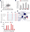
(A) Alanine scanning mutagenesis of EBOV GP. Clones with <25% binding compared to wild-type (WT) EBOV GP yet >65% reactivity for a control mAb were considered critical for CA45 binding (red). (B) Mutations of four individual residues reduced CA45 binding (red bars) but retained FVM04 and FVM09 binding (gray bars). Bars represent the mean and range of at least two replicates. (C) Homology between filovirus GP sequences within the regions encompassing the CA45 epitope. Conserved residues in blue and CA45 critical residues in red. Arrows correspond to β strands in EBOV GP structure. (D) Position of GP residues critical for CA45 in the structure of trimeric EBOV GP (dashed circle). (E) Binding of CA45 to selected EBOV GP mutants determined by flow cytometry. MFI: mean fluorescence intensity. Also see Tables S5 and S6, and Figures S4 and S5.
Figure 5
Figure 5. Single-particle EM analysis of CA45 Fab bound to EBOV GPΔMuc
(A) 3D EM reconstruction of CA45 Fab bound to EBOV GPΔMuc (gray) with EBOV GPΔMuc structure (light blue, PDB 5JQ3) docked in. (B) The same EM map with a single EBOV GP protomer docked in, with the internal fusion loop (IFL) highlighted in dark blue. (C) LC (orange) and HC (purple) of CA45 Fab homology model docked into EM map. The CDRH1 and CDRH3 of CA45 were docked in close proximity to GP, based on the CA45 CDR ala scanning data. R64, Y517, G546, and N550 that are critical for CA45 binding are highlighted in dark blue spheres, K190 in light blue spheres. (D) Comparison of CA45 and KZ52 epitopes. Upper, bottom view of the EM map with KZ52 Fab bound to EBOV GP protomer (PDB 5HJ3) with the epitope highlighted in red. Middle, the CA45 Fab epitope (purple) includes overlapping residues (yellow) with the KZ52 epitope. Lower, The GP residues contacted by KZ52 are less conserved among ebolaviruses compared to CA45. (E) Comparison of Ebola GP interactions with CA45 and EBOV-specific neutralizing mAbs (KZ52, c2G4, and c4G7) at the GP1-GP2 interface. Left, top view. Right, side view. c2G4 Fab in yellow (EMD-6151), c4G7 Fab in purple (EMD-6152). KZ52 (PDB 5HJ3, blue) also contacts similar residues as c2G4. CA45 Fab (gray) has significant overlap with these three EBOV antibodies, but with its shifted angle of approach, may confer breadth of neutralization. Also see Figure S6.
Figure 6
Figure 6. Efficacy in mouse and guinea pig models
(A) Groups of 10 or 20 BALB/c mice were infected with 100 pfu of MA-EBOV and treated with an IP injection of CA45 at indicated doses or PBS at 2 dpi and monitored for 21 days. p values for each treatment group compared to the PBS group was determined by Log-rank (Mantel-Cox) test. (B) IFNAR−/− mice were infected with 1000 pfu of SUDV and treated at 1 dpi with an IP injection of either PBS, FVM04, CA45, or FVM04+CA45 at indicated doses, and monitored for 21 days. (C) Hartley guinea pigs were infected with 1000 LD50 of GPA-EBOV and treated at 3 dpi by IP injection of FVM04, CA45, the cocktail (n=6), or PBS (n=4) and monitored for 28 days. p values: CA45 vs. PBS, p =0.0018; FVM04 vs. PBS, p=0.0018; cocktails vs. PBS p<0.0001; CA45 vs. cocktails, p=0.0079; FVM04 vs. cocktails, p<0.0001. (D) Guinea pigs were challenged with GPA-SUDV and treated with 5 mg of CA45 (n=6) or PBS (n=4) at 3 dpi and monitored for 28 days, p<0.0001.
Figure 7
Figure 7. Efficacy in ferret model of BDBV infection
Groups of two male and two female ferrets (denoted with M or F suffix to animal number) were infected with 253 TCID50 of BDBV followed by IP treatment at 3 and 6 dpi with 20 mg each of CA45 and FVM04. Two control animals (780M and 727F) received PBS only. Protection data are shown as percent survival (A), clinical score (B), weight change (C), viral burden in blood (D), and tissue swabs (E–G), as well as blood chemistry markers (H-L). Also see Figure S7.

Comment in

References

    1. Bornholdt ZA, Ndungo E, Fusco ML, Bale S, Flyak AI, Crowe JE, Jr, Chandran K, Saphire EO. Host-Primed Ebola Virus GP Exposes a Hydrophobic NPC1 Receptor-Binding Pocket, Revealing a Target for Broadly Neutralizing Antibodies. MBio. 2016a;7:e02154–02115. - PMC - PubMed
    1. Bornholdt ZA, Turner HL, Murin CD, Li W, Sok D, Souders CA, Piper AE, Goff A, Shamblin JD, Wollen SE, et al. Isolation of potent neutralizing antibodies from a survivor of the 2014 Ebola virus outbreak. Science. 2016b;351:1078–1083. - PMC - PubMed
    1. Brannan JM, Froude JW, Prugar LI, Bakken RR, Zak SE, Daye SP, Wilhelmsen CE, Dye JM. Interferon alpha/beta Receptor-Deficient Mice as a Model for Ebola Virus Disease. J Infect Dis 2015 - PubMed
    1. Brister JR, Bao Y, Zhdanov SA, Ostapchuck Y, Chetvernin V, Kiryutin B, Zaslavsky L, Kimelman M, Tatusova TA. Virus Variation Resource–recent updates and future directions. Nucleic Acids Res. 2014;42:D660–665. - PMC - PubMed
    1. Carroll MW, Matthews DA, Hiscox JA, Elmore MJ, Pollakis G, Rambaut A, Hewson R, Garcia-Dorival I, Bore JA, Koundouno R, et al. Temporal and spatial analysis of the 2014–2015 Ebola virus outbreak in West Africa. Nature. 2015;524:97–101. - PMC - PubMed

MeSH terms