. 2017 May 19;10(1):112.
doi: 10.1186/s13045-017-0435-x.

Targeting the CXCR4 pathway using a novel anti-CXCR4 IgG1 antibody (PF-06747143) in chronic lymphocytic leukemia

Affiliations

Targeting the CXCR4 pathway using a novel anti-CXCR4 IgG1 antibody (PF-06747143) in chronic lymphocytic leukemia

Manoj K Kashyap et al. J Hematol Oncol. .

Abstract

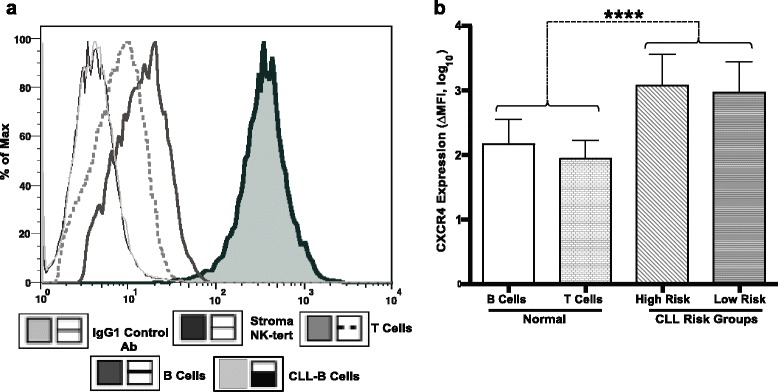
Background: The CXCR4-CXCL12 axis plays an important role in the chronic lymphocytic leukemia (CLL)-microenvironment interaction. Overexpression of CXCR4 has been reported in different hematological malignancies including CLL. Binding of the pro-survival chemokine CXCL12 with its cognate receptor CXCR4 induces cell migration. CXCL12/CXCR4 signaling axis promotes cell survival and proliferation and may contribute to the tropism of leukemia cells towards lymphoid tissues and bone marrow. Therefore, we hypothesized that targeting CXCR4 with an IgG1 antibody, PF-06747143, may constitute an effective therapeutic approach for CLL.

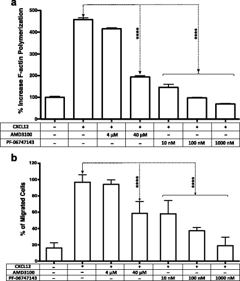
Methods: Patient-derived primary CLL-B cells were assessed for cytotoxicity in an in vitro model of CLL microenvironment. PF-06747143 was analyzed for cell death induction and for its potential to interfere with the chemokine CXCL12-induced mechanisms, including migration and F-actin polymerization. PF-06747143 in vivo efficacy was determined in a CLL murine xenograft tumor model.

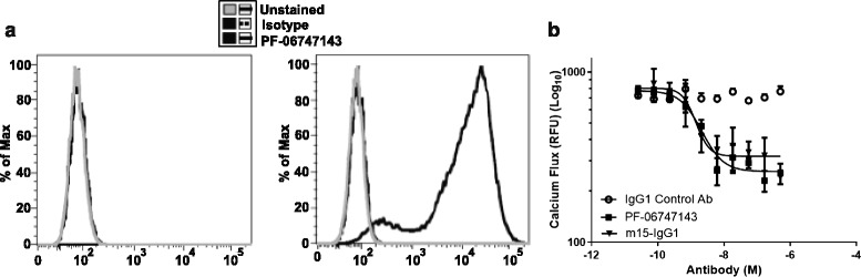
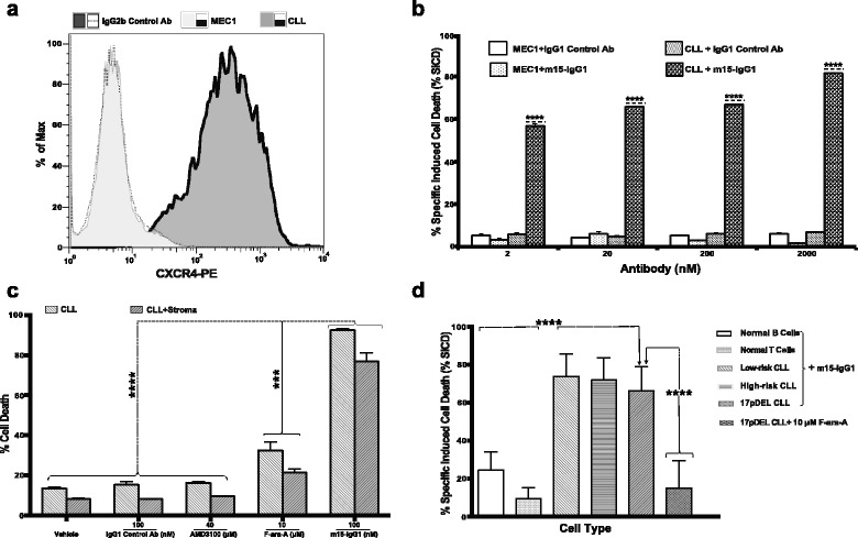
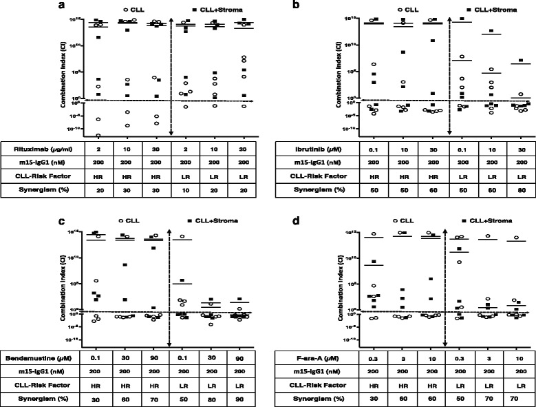
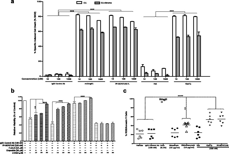
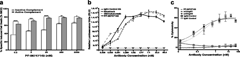
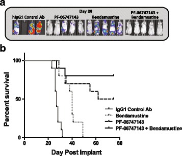
Results: PF-06747143, a novel-humanized IgG1 CXCR4 antagonist antibody, induced cell death of patient-derived primary CLL-B cells, in presence or absence of stromal cells. Moreover, cell death induction by the antibody was independent of CLL high-risk prognostic markers. The cell death mechanism was dependent on CXCR4 expression, required antibody bivalency, involved reactive oxygen species production, and did not require caspase activation, all characteristics reminiscent of programmed cell death (PCD). PF-06747143 also induced potent B-CLL cytotoxicity via Fc-driven antibody-dependent cell-mediated cytotoxicity (ADCC) and complement-dependent cytotoxicity activity (CDC). PF-06747143 had significant combinatorial effect with standard of care (SOC) agents in B-CLL treatment, including rituximab, fludarabine (F-ara-A), ibrutinib, and bendamustine. In a CLL xenograft model, PF-06747143 decreased tumor burden and improved survival as a monotherapy, and in combination with bendamustine.

Conclusions: We show evidence that PF-06747143 has biological activity in CLL primary cells, supporting a rationale for evaluation of PF-06747143 for the treatment of CLL patients.

Keywords: ADCC; CDC; CXCL12; CXCR4; Cell death; Chemokine; Chronic lymphocytic leukemia; PF-06747143; Reactive oxygen species.


Figures

Fig. 1
Fig. 1
Expression of CXCR4 receptor in CLL-B, normal B, T, and stroma-NK-tert cells. a CXCR4 expression was assessed by surface staining using an anti-CXCR4 antibody in CLL-B, normal B, T, and stroma-NK-tert cells. A representative panel is shown. b CXCR4 expression was evaluated by flow cytometry in CLL patient cells with high-risk and low-risk characteristics (n = 20 per group) and in normal B and T cells obtained from healthy donors (n = 5 per group). The figure shows the mean fluorescence intensities (MFI) ± standard deviation (SD) of the samples analyzed in duplicate for CXCR4 expression in each cell type. Statistical significance was determined by using Bonferroni’s correction test for multiple comparison tests, where *, **, ***, and **** represent p < 0.05; p < 0.01, p < 0.001, and p < 0.0001, respectively
Fig. 2
Fig. 2
PF-06747143 binds specifically to human CXCR4-expressing cells and blocks CXCL12-induced calcium flux. a CHO-parental and CHO-hCXCR4 cell lines were exposed to 20 μg/mL of either a human IgG1 ĸ-PE antibody (isotype control) or PF-06747143-PE and analyzed by flow cytometry. b Calcium flux assay was performed in human T cell leukemia Jurkat cells incubated with PF-06747143, m15-IgG1, or isotype control IgG1 antibody in presence of CXCL12 at 8 nM. Experiment was performed in quadruplicates. Shown are mean intracellular calcium concentrations in relative fluorescence units (RFU). ± standard error of the mean (SEM)
Fig. 3
Fig. 3
CXCR4 antibody-induced cell death is dependent on CXCR4-expression and independent of CLL disease risk factor or stromal presence. a CXCR4 expression profiling was done using an anti-CXCR4 antibody for staining in the MEC1 cell line and primary CLL-B cells from a representative patient, followed by analysis using flow cytometry. The CXCR4 expression is presented in ∆MFI. b MEC1 and CLL-B cells were treated with different concentrations of m15-IgG1 (2–2000 nM) or IgG1 control antibody, for 48 h followed by flow cytometry analysis to determine % SICD. Samples were tested in duplicates, with the mean and standard deviation shown for each group. c The CLL-B cells derived from a CLL patient were treated with either F-ara-A (3 and 10 μM), AMD3100 (4 and 40 μM), IgG1 control antibody, or m15-IgG1 antibody, in presence or absence of stroma NK-tert cells, for 48 h followed by analysis using flow cytometry. The results of samples analyzed in duplicates with the mean ± SD are shown for each group. d Primary leukemia CLL-B cells were obtained from CLL patients, with high-risk or low-risk phenotypes, or carrying TP53 17pDel mutation (n = 10 per group). Normal B and T lymphocytes were obtained from healthy donors (n = 10 per group). The % SICD was determined after treatment with 100 nM of m15-IgG1 for 48 h. The figure shows the mean ± SD for % SICD in different cell types. Statistical significance was determined using Bonferroni’s correction test for multiple comparison tests, where *, **, ***, and **** represent p < 0.05; p < 0.01, p < 0.001, and p < 0.0001, respectively
Fig. 4
Fig. 4
Cell death synergism of m15-IgG1 antibody with components of standard of care (SOC) in CLL. The primary leukemia CLL-B cells were incubated either alone or with stroma-NK-tert cells and treated with m15-IgG1 (200 nM) and three different concentrations of SOC agents a rituximab (1, 3, and 10 μg/mL), b ibrutinib (0.1, 10, and 30 μM), c bendamustine (0.1, 30, and 90 μM), and d fludarabine (F-ara-A, 1, 3, and 10 μM). Treatments were performed with each agent alone or in combination. The % cell death was used to calculate the median effect of the combinatorial effect of m15-IgG1 with each different SOC agent. The synergism between m15-IgG1 and SOCs was expressed as a combination index (CI), which uses the definitions: additive (CI = 1), synergistic (CI < 1), and antagonistic (CI > 1). The data were analyzed. The empty circle symbols (O) denote CLL cells alone, while solid black symbols (■) denote CLL cells co-cultured with stroma-NK-tert cells
Fig. 5
Fig. 5
PF-06747143-induced cell death is bivalency-dependent, involves ROS, but not caspase activation. a Primary leukemia B cells obtained from a CLL patient were cultured alone or in presence of stroma-NK-tert cells after treatment with 10, 100, and 1000 nM of m15-IgG1, PF-06747143, or its Fab and F(ab’)2 forms, for 48 h. % SICD was measured by flow cytometry using CD19/CD5/Annexin V staining. The results of samples analyzed in duplicates with the mean ± SD are shown for each group. b CLL cells were incubated with PF-06747143 (100 nM), F-ara-A (10 μM), or etoposide (30 μM) for 48 h, either alone or in combination with different concentrations of a pan-caspase inhibitor, Z-VAD-FMK (Z-VAD) (10, 30, 90 μM). CLL cell death was analyzed by flow cytometry. c CLL-B cells (n = 6 per group) were incubated for 48 h with the intact CXCR4 antibody PF-06747143, the Fab and the F(ab’)2 forms of the antibody, the CD20 antibodies rituximab or obinutuzumab, or the IgG1 control antibody. H2O2 was used as a positive control. ROS production and cell dealth were analyzed by co-staining for ROS and CD19+/CD5+/Annexin V, respectively. The figure shows the individual data points for each group, and horizontal lines represent the mean of each group. Statistical significance was determined using Bonferroni’s correction test where *, **, ***, and **** represent p < 0.05; p < 0.01, p < 0.001, and p < 0.0001, respectively
Fig. 6
Fig. 6
PF-06747143 inhibits CXCL12-induced tumor cell actin polymerization and migration. a B-CLL patient cells were treated with no compound (negative control), AMD3100 (4 and 40 μM), or PF-06747143 (10, 100, and 1000 nM) prior to stimulation with CXCL12 (90 nM) for 15 s. F-actin polymerization was measured using FITC-labeled phalloidin in CD19/CD5-pre-labeled CLL patient cells. All samples are plotted relative to the mean fluorescence intensity of the negative control group, without chemokine CXCL12, set to 100%. The results of samples analyzed in duplicates with the mean ± SD are shown for each group. b CLL patient primary cells were incubated with PF-06747143 (10, 100, 1000 nM) or AMD3100 (4 and 40 μM) for 1 h and loaded onto a transwell chamber and incubated for 2 h in the presence of CXCL12 (12 nM) or media control. Cells that migrated to the lower chamber were enumerated using flow cytometry. The results of samples analyzed in duplicates with the mean ± SD are shown for each group. Statistical significance was determined using Bonferroni’s correction test
Fig. 7
Fig. 7
PF-06747143 induces CLL-B cell killing by ADCC and CDC. a CDC assay was performed by treating CLL-B cells (1 × 106/mL) with PF-06747143, in presence of complete or heat-inactivated 5% human serum. The cells were incubated for 4 h and the cytotoxicity was determined using flow cytometry with CD19/CD5/Annexin V staining. b The ADCC assay in patient B-CLL cells was performed using 1:1 ratio of the target/effector cell (T/E) and incubated for 6 h at 37 °C. The IgG1 control antibody, PF-06747143, and rituximab were tested in a 1:3 titration curve, ADCC activity was determined using Bio-Glo™ luciferase assay, and the luminescence results are expressed in relative light units (RLU). The samples were analyzed in duplicates with the mean and SD shown for each group. The data was analyzed using Prism 4 GraphPad software. c ADCC activity was evaluated in JVM-13 tumor cells by incubating the cells with PF-06747143, m15-IgG1, m15-IgG4, or respective negative control antibodies for 4 h, in the presence of NK92 158V effector killer cells (effector/target cell ratio 10:1). Cell lysis was measured by ToxiLight bioluminescent cytotoxicity assay. Experiments were performed in quadruplicates with the mean ± SEM shown for each group
Fig. 8
Fig. 8
PF-06747143 inhibits tumor burden and increases survival as a monotherapy or in combination with bendamustine, in a disseminated CLL tumor model. JVM-13-Luc CLL cells were implanted intravenously (1 × 106 cells) and allowed to spontaneously migrate and home in the bone marrow and lymph nodes for 19 days, when animals were randomized (n = 10/group). Animals were treated with IgG1 control or PF-06747143 antibodies at 10 mg/kg, subcutaneously, weekly, for six doses. Bendamustine was dosed at 30 mg/kg, intraperitoneally, on days 19 and 20, followed by another 2-day cycle 28 days later a Whole-body bioluminescence representative imaging showing bone marrow tumor burden on day 26. b Kaplan-Meier survival curve, using hind leg paralysis as endpoint

Similar articles

Cited by

References

    1. Keshava Prasad TS, Goel R, Kandasamy K, Keerthikumar S, Kumar S, Mathivanan S, Telikicherla D, Raju R, Shafreen B, Venugopal A, et al. Human Protein Reference Database—2009 update. Nucleic Acids Res. 2009;37(Database issue):D767–72. doi: 10.1093/nar/gkn892. - DOI - PMC - PubMed
    1. Lee B, Sharron M, Montaner LJ, Weissman D, Doms RW. Quantification of CD4, CCR5, and CXCR4 levels on lymphocyte subsets, dendritic cells, and differentially conditioned monocyte-derived macrophages. Proc Natl Acad Sci U S A. 1999;96(9):5215–20. doi: 10.1073/pnas.96.9.5215. - DOI - PMC - PubMed
    1. Murdoch C. CXCR4: chemokine receptor extraordinaire. Immunol Rev. 2000;177:175–84. doi: 10.1034/j.1600-065X.2000.17715.x. - DOI - PubMed
    1. Burger M, Hartmann T, Krome M, Rawluk J, Tamamura H, Fujii N, Kipps TJ, Burger JA. Small peptide inhibitors of the CXCR4 chemokine receptor (CD184) antagonize the activation, migration, and antiapoptotic responses of CXCL12 in chronic lymphocytic leukemia B cells. Blood. 2005;106(5):1824–30. doi: 10.1182/blood-2004-12-4918. - DOI - PubMed
    1. Teicher BA, Fricker SP. CXCL12 (SDF-1)/CXCR4 pathway in cancer. Clin Cancer Res. 2010;16(11):2927–31. doi: 10.1158/1078-0432.CCR-09-2329. - DOI - PubMed

Publication types

MeSH terms