. 2017 Aug 1;357(1):116-123.
doi: 10.1016/j.yexcr.2017.05.009. Epub 2017 May 17.

Small-molecule inhibition of Wnt signaling abrogates dexamethasone-induced phenotype of primary human trabecular meshwork cells

Affiliations

Small-molecule inhibition of Wnt signaling abrogates dexamethasone-induced phenotype of primary human trabecular meshwork cells

Sarah D Ahadome et al. Exp Cell Res. .

Abstract

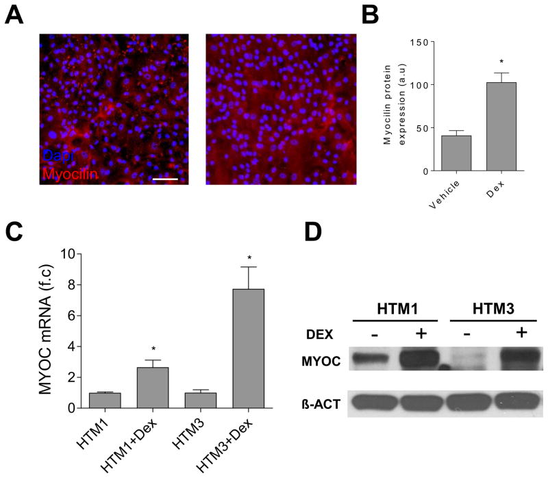
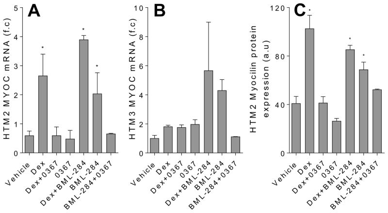
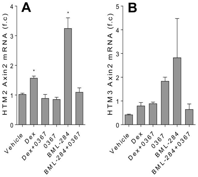
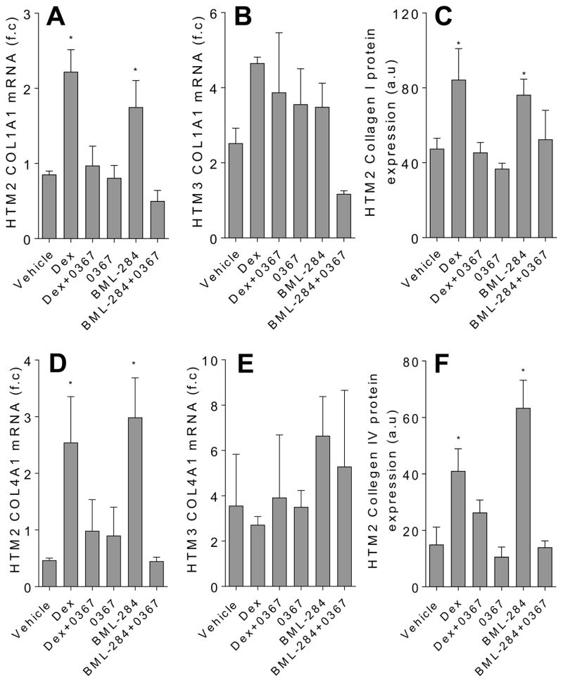
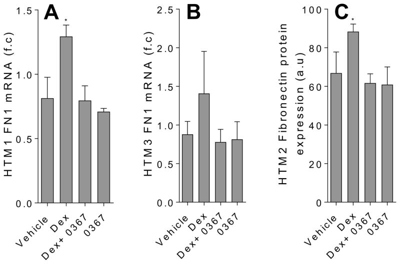
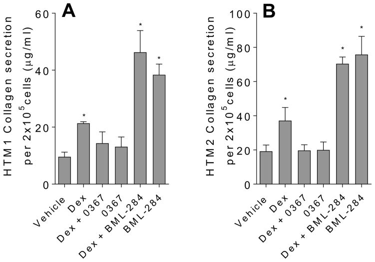
Trabecular meshwork (TM) cells are the governing regulators of the TM structure. When the functionality of these cells is impaired, the structure of the TM is perturbed which often results in increased ocular hypertension. High intraocular pressure is the most significant risk factor for steroid-induced glaucoma. Dexamethasone (Dex)-induced phenotype of TM cells is widely utilized as a model system to gain insight into mechanisms underlying damaged TM in glaucoma. In this study, to assess the possible role of abnormal Wnt signaling in steroid-induced glaucoma, we analyzed the effects of small-molecule Wnt signaling modulators on Dex-induced expression extracellular matrix proteins of primary human TM cells. While Dex-treated TM cells exhibited increased collagen and fibronectin expression, we found that Wnt signaling inhibitor 3235-0367 suppressed these Dex-induced effects. We therefore propose that Wnt signaling plays an important role in Dex-mediated impairment of TM cell functions. Moreover, the use of small-molecule Wnt signaling inhibitors to treat TM cells may provide an opportunity of restoring TM tissue in steroid-induced glaucoma.

Keywords: Dexamethasone; Glaucoma; Trabecular meshwork cell; Wnt.


Conflict of interest statement

Conflicts of interest

The authors declare that they have no conflicts of interest with any of the content in this article except that J.S. is an employee of ScienCell Research Laboratories.

Figures

Figure 1
Figure 1
TM cell characterization. Representative immunofluorescence images (A) and the summary of the immunofluorescence study (B) showed that the myocilin level in the HTM2 cells was increased after Dex treatment (100 nM) for seven days. (C) The myocilin mRNA levels elevated in both the HTM1 cells and the HTM3 cells after Dex (100 nM) treatment for five days. Each experiment was carried out at two independent times; each measurement was done in triplicate. (D) The Western Blot studies showed that the myocilin protein levels increased in both HTM1 and HTM3 after Dex treatment for five days.
Figure 2
Figure 2
Wnt signaling inhibition decreases Dex-mediated myocilin mRNA and protein expression in TM cells. Analysis of myocilin mRNA expression using qPCR with f.c stands for fold change in HTM2 (A) and HTM3 (B), as well as myocilin protein expression in HMT2 using immunofluorescence where a.u stands for arbitrary units (C). The cells were treated for 7 days with DMSO vehicle (0.002 %), Dex (100 nM), Dex in the presence of Wnt signaling activator BML-284 (0.1 μM), BML-284 alone (0.1 μM), Dex in the presence of Wnt signaling inhibitor 3235-0367 (1 μM), 3235-0367 alone (1 μM) or lastly BML-284 (0.1 μM) together with 3235-0367 (1 μM). The mRNA expression data were obtained from three independent experiments (* p<0.05 via One-way ANOVA with Dunnett’s correction) and the raw immunofluorescence images are shown in the Supplementary figure S1.
Figure 3
Figure 3
Dex-induced AXIN2 expression in TM cells is decreased with Wnt signaling inhibition. Analysis of AXIN2 mRNA expression was carried out using qPCR. The HTM2 (A) and HTM3 (B) cells were treated for 7 days with DMSO vehicle (0.002 %), Dex (100 nM), Dex in the presence of Wnt signaling inhibitor 3235-0367 (1 μM), 3235-0367 alone (1 μM), Wnt signaling activator BML-284 (0.1 μM) or BML-284 (0.1 μM) together with 3235-0367 (1 μM). The data were obtained from three independent experiments (* p<0.05 via One-way ANOVA with Dunnett’s correction).
Figure 4
Figure 4
Inhibition of Wnt signaling decreases the expression of collagen in the Dex-treated TM cells. The top panel shows the analysis of collagen I (COL1A1) mRNA in HTM2 (A) and HTM3 (B) cells and protein expression in the HTM2 cells (C) using qPCR and immunofluorescence respectively. The bottom panel shows the analysis of collagen IV (COL4A1) mRNA in the HTM2 (D) and HTM3 (E) cells and protein expression in the HTM2 cells (F). The cells were treated for 7 days with DMSO vehicle (0.002 %), Dex (100 nM), Dex in the presence of Wnt signaling inhibitor 3235-0367 (1 μM), 3235-0367 alone (1 μM), Wnt signaling activator BML-284 (0.1 μM) or BML-284 (0.1 μM) together with 3235-0367 (1 μM). Each experimental condition was repeated three times (* p<0.05 via One-way ANOVA with Dunnett’s correction). The raw immunofluorescent images are shown in the Supplementary figure S2.
Figure 5
Figure 5
Inhibition of Wnt signaling decreases the expression fibronectin the in Dex-treated TM cells. The analysis of fibronectin (FN1) mRNA in HTM2 (A) and HTM3 (B) cells and protein expression in HTM2 cells (C) were carried out using qPCR and immunofluorescence respectively. The cells were treated for 7 days with DMSO vehicle (0.002 %), Dex (100 nM), Dex in the presence of Wnt signaling inhibitor 3235-0367 (1 μM), and 3235-0367 alone (1 μM). Each experimental condition was repeated three times (* p<0.05 via One-way ANOVA with Dunnett’s correction). The raw immunofluorescent images are shown in the Supplementary figure S3.
Figure 6
Figure 6
Inhibition of Wnt signaling reduces collagen secretion from TM cells treated with Dex. The analysis of collagen secreted in the conditioned media were collected from HTM2 (A) and HTM3 (B) using the Sircol method of collagen detection. Cells were treated with the following conditions: ethanol (vehicle) (0.004%), Dex (100 nM), Dex (100 nM) in the presence of Wnt signaling inhibitor 3235-0367 (1 μM), 3235-0367 alone (1 μM), Dex (100 nM) in the presence of Wnt signaling activator BML-284 (0.1 μM), and activator BML-284 alone (0.1 μM). Each experiment condition was carried out in triplicates (*p<0.05 via One way ANOVA with Dunnett’s correction).

References

    1. Armaly MF. Effect of Corticosteroids on Intraocular Pressure and Fluid Dynamics. Ii. The Effect of Dexamethasone in the Glaucomatous Eye. Arch Ophthalmol. 1963;70:492–499. - PubMed
    1. Cubey RB. Glaucoma following the application of corticosteroid to the skin of the eyelids. Br J Dermatol. 1976;95:207–208. - PubMed
    1. Pleyer U, Ursell PG, Rama P. Intraocular Pressure Effects of Common Topical Steroids for Post-Cataract Inflammation: Are They All the Same? Ophthalmology and Therapy. 2013;2:55–72. - PMC - PubMed
    1. Junglas B, Yu AH, Welge-Lussen U, Tamm ER, Fuchshofer R. Connective tissue growth factor induces extracellular matrix deposition in human trabecular meshwork cells. Exp Eye Res. 2009;88:1065–1075. - PubMed
    1. Pattabiraman PP, Rao PV. Mechanistic basis of Rho GTPase-induced extracellular matrix synthesis in trabecular meshwork cells. Am J Physiol Cell Physiol. 2010;298:C749–763. - PMC - PubMed

Publication types

MeSH terms