. 2017 Aug 1;313(2):H265-H274.
doi: 10.1152/ajpheart.00752.2016. Epub 2017 May 19.

Acetylation of mitochondrial proteins by GCN5L1 promotes enhanced fatty acid oxidation in the heart

Affiliations

Acetylation of mitochondrial proteins by GCN5L1 promotes enhanced fatty acid oxidation in the heart

Dharendra Thapa et al. Am J Physiol Heart Circ Physiol. .

Abstract

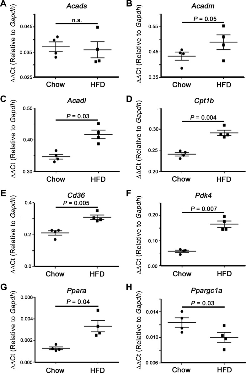
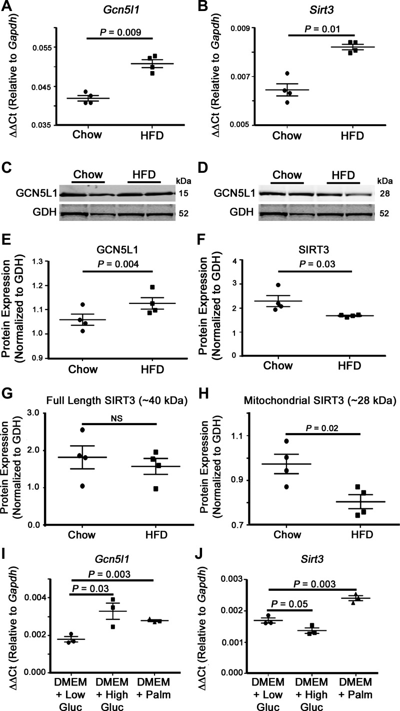
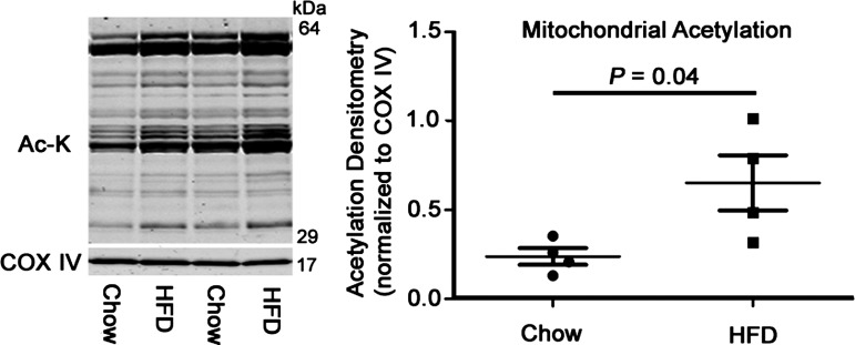
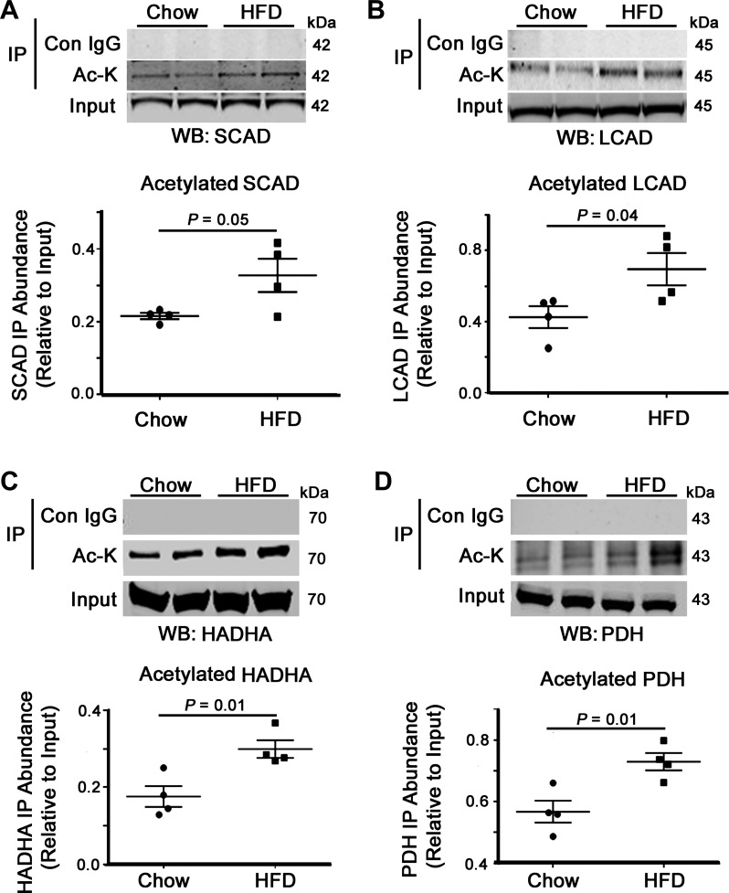
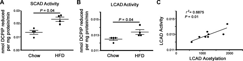
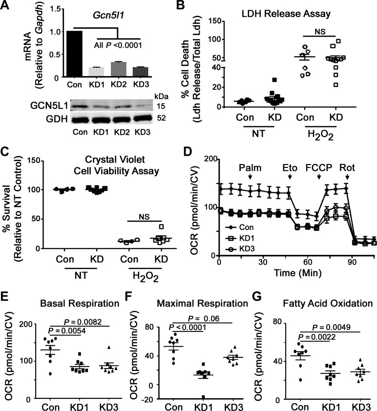
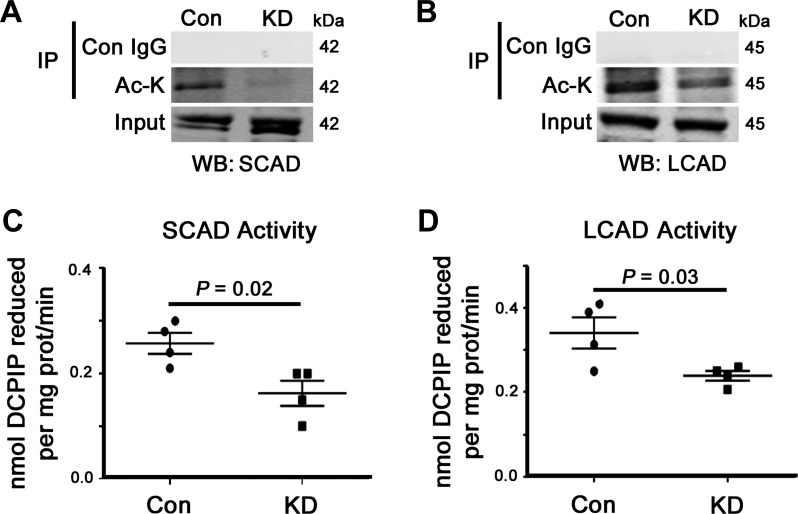
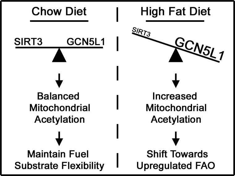
Lysine acetylation is a reversible posttranslational modification and is particularly important in the regulation of mitochondrial metabolic enzymes. Acetylation uses acetyl-CoA derived from fuel metabolism as a cofactor, thereby linking nutrition to metabolic activity. In the present study, we investigated how mitochondrial acetylation status in the heart is controlled by food intake and how these changes affect mitochondrial metabolism. We found that there was a significant increase in cardiac mitochondrial protein acetylation in mice fed a long-term high-fat diet and that this change correlated with an increase in the abundance of the mitochondrial acetyltransferase-related protein GCN5L1. We showed that the acetylation status of several mitochondrial fatty acid oxidation enzymes (long-chain acyl-CoA dehydrogenase, short-chain acyl-CoA dehydrogenase, and hydroxyacyl-CoA dehydrogenase) and a pyruvate oxidation enzyme (pyruvate dehydrogenase) was significantly upregulated in high-fat diet-fed mice and that the increase in long-chain and short-chain acyl-CoA dehydrogenase acetylation correlated with increased enzymatic activity. Finally, we demonstrated that the acetylation of mitochondrial fatty acid oxidation proteins was decreased after GCN5L1 knockdown and that the reduced acetylation led to diminished fatty acid oxidation in cultured H9C2 cells. These data indicate that lysine acetylation promotes fatty acid oxidation in the heart and that this modification is regulated in part by the activity of GCN5L1.NEW & NOTEWORTHY Recent research has shown that acetylation of mitochondrial fatty acid oxidation enzymes has greatly contrasting effects on their activity in different tissues. Here, we provide new evidence that acetylation of cardiac mitochondrial fatty acid oxidation enzymes by GCN5L1 significantly upregulates their activity in diet-induced obese mice.

Keywords: GCN5L1; acetylation; fatty acid oxidation; heart; high-fat diet; mitochondria; sirtuin 3.


Conflict of interest statement

No conflicts of interest, financial or otherwise, are declared by the authors.

Figures

Fig. 1.
Fig. 1.
Expression of cardiac fuel metabolism genes. High-fat diet (HFD) promotes an enhanced fatty acid oxidation phenotype in the heart. A and B: no significant difference was observed in short-chain acyl-CoA dehydrogenase (Acads, SCAD) or medium-chain acyl-CoA dehydrogenase (Acadm, MCAD) levels. C–G: significant increases in the mRNA content of long-chain acyl-CoA dehydrogenase (Acadl, LCAD), carnitine palmitoyltransferase 1b (Cpt1b), Cd36, pyruvate dehydrogenase kinase 4 (Pdk4), and peroxisome proliferator-activted receptor-α (Ppara) were observed with HFD feeding, whereas peroxisome proliferator-activated receptor-γ coactivator-1α (Ppargc1a; H) was significantly decreased. Values are expressed as means ± SE; n = 4 per group. P value significance is as shown in the graphs using a two-way Student’s t-test.
Fig. 2.
Fig. 2.
Expression of mitochondrial acetylation regulators in cardiac mitochondria. HFD feeding led to a proacetylation phenotype in cardiac mitochondrial proteins. A and B: mitochondrial acetyltransferase Gcn5l1 and deacetylase sirtuin 3 (Sirt3) mRNA expression were significantly increased in cardiac mitochondria from chronic HFD-fed mice. C–E: whereas the GCN5L1 protein level was significantly increased in HFD-fed animals, SIRT3 protein abundance was significantly decreased. Western blots for GCN5L1 and SIRT3 were obtained from the same gel, and therefore the same GDH loading control was used for each blot. G: there was no significant difference in the abundance of the 40-kDa, preimport, cytosolic form of SIRT3 in chow- and HFD-fed mice. H: in contrast, there was a significant decrease in the 28-kDa mitochondrial form of the enzyme in HFD-fed animals. I and J: Gcn5l1 gene expression in H9C2 cells was significantly increased after 4 h of high glucose (25 mM) and palmitate (200 μM) exposure in basal DMEM relative to low glucose (5 mM) exposure, whereas Sirt3 expression was only elevated in response to palmitate. Values are expressed as means ± SE; n = 4 per group. P value significance is as shown in the graphs using two-way Student’s t-test.
Fig. 3.
Fig. 3.
Global mitochondrial lysine acetylation in chow- and HFD-fed hearts. Overall cardiac mitochondrial protein acetylation was significantly increased, with no changes observed in the protein content of cytochrome c oxidase subunit 4 (COX IV). Values are expressed as means ± SE; n = 4 per group. P value significance is as shown in the graphs using two-way Student’s t-test.
Fig. 4.
Fig. 4.
Impact of HFD on metabolic enzyme acetylation status. HFD feeding led to increased fatty acid oxidation (FAO) enzyme acetylation, which contributes to upregulated fatty acid utilization in diet-induced obese mice. A–C: the acetylated lysine pulldown showed an increase in the acetylation levels of the FAO proteins SCAD, LCAD, and hydroxyacyl-CoA dehydrogenase (HADHA) and the pyruvate oxidation enzyme pyruvate dehydrogenase (PDH; D) from HFD-fed cardiac tissue relative to chow-fed controls. Values are expressed as means ± SE; n = 4 per group. P value significance is as shown in the graphs using two-way Student’s t-test.
Fig. 5.
Fig. 5.
Impact of HFD-related acetylation on in vitro FAO enzyme activity. A and B: enzymatic activity of SCAD and LCAD was significantly elevated in cardiac tissues from HFD-fed mice. C: regression analysis showed a significant correlation between LCAD acetylation status and enzymatic activity from all tested mice. Values are expressed as means ± SE, n = 4 per group. P value significance is as shown in the graphs using two-way Student’s t-test.
Fig. 6.
Fig. 6.
Regulation of cellular respiration and FAO by GCN5L1. GCN5L1 is a key regulator of FAO in cardiac cells. A: mRNA and protein levels of three stable GCN5L1 knockdown H9C2 cell lines created using different lentivirus-derived shRNAs. Glutamate dehydrogenase (GDH) was used as the loading control. Significant decreases in both mRNA and protein content were observed in two lines (KD1 and KD3). B and C: there was no significant difference in either cell death (LDH release) or cell viability (crystal violet staining) in GCN5L1 knockdown cells relative to the control when exposed to 50 mM H2O2 for 30 min. D: mitochondrial bioenergetic profile of GCN5L1 control, KD1, and KD3 cells after the addition of palmitate (Palm), etomoxir (Eto), carbonyl cyanide-4-(trifluoromethoxy) phenylhydrazone (FCCP), and rotenone (Rot). E–G: there was a significant decrease in basal respiration, maximal uncoupled respiration (for KD1), and FAO in GCN5L1 knockdown cells relative to the control. Values are expressed as means ± SE; n = 4 per group for GCN5L1 mRNA and protein levels and n = 8 per group for respiration measurements. P value significance is as shown in the graphs. One-way ANOVA was used for A and E−G; two-way ANOVA was used for B and C; Tukey post hoc testing was performed in each case.
Fig. 7.
Fig. 7.
Impact of GCN5L1 knockdown on cardiac FAO enzyme acetylation and activity. GCN5L1 promoted the acetylation and activity of cardiac FAO proteins. A and B: our immunoprecipitation-based acetylation assay showed a large decrease in the acetylation status of SCAD and LCAD proteins in GCN5L1 knockdown cells relative to the control. C and D: this decrease correlated with reduced enzymatic activities of SCAD and LCAD in vitro. Values are expressed as means ± SE; n = 4 per group. P value significance is as shown in the graphs using two-way Student’s t-test.
Fig. 8.
Fig. 8.
Model of lysine acetylation and FAO in cardiac metabolism. Under normal conditions, a balance between lysine acetylation and deacetylation allows mitochondria to maintain bioenergetic output under changing physiological conditions. In obese mice, increased acetylation of mitochondrial proteins drives a pro-FAO metabolic phenotype, thereby reducing substrate flexibility in the heart.

Comment in

References

    1. Alrob OA, Sankaralingam S, Ma C, Wagg CS, Fillmore N, Jaswal JS, Sack MN, Lehner R, Gupta MP, Michelakis ED, Padwal RS, Johnstone DE, Sharma AM, Lopaschuk GD. Obesity-induced lysine acetylation increases cardiac fatty acid oxidation and impairs insulin signalling. Cardiovasc Res 103: 485–497, 2014. doi: 10.1093/cvr/cvu156. - DOI - PMC - PubMed
    1. Anderson KA, Hirschey MD. Mitochondrial protein acetylation regulates metabolism. Essays Biochem 52: 23–35, 2012. doi: 10.1042/bse0520023. - DOI - PMC - PubMed
    1. Bakermans AJ, Dodd MS, Nicolay K, Prompers JJ, Tyler DJ, Houten SM. Myocardial energy shortage and unmet anaplerotic needs in the fasted long-chain acyl-CoA dehydrogenase knockout mouse. Cardiovasc Res 100: 441–449, 2013. doi: 10.1093/cvr/cvt212. - DOI - PubMed
    1. Banks AS, Kon N, Knight C, Matsumoto M, Gutiérrez-Juárez R, Rossetti L, Gu W, Accili D. SirT1 gain of function increases energy efficiency and prevents diabetes in mice. Cell Metab 8: 333–341, 2008. doi: 10.1016/j.cmet.2008.08.014. - DOI - PMC - PubMed
    1. Bharathi SS, Zhang Y, Mohsen AW, Uppala R, Balasubramani M, Schreiber E, Uechi G, Beck ME, Rardin MJ, Vockley J, Verdin E, Gibson BW, Hirschey MD, Goetzman ES. Sirtuin 3 (SIRT3) protein regulates long-chain acyl-CoA dehydrogenase by deacetylating conserved lysines near the active site. J Biol Chem 288: 33837–33847, 2013. doi: 10.1074/jbc.M113.510354. - DOI - PMC - PubMed

MeSH terms

LinkOut - more resources