. 2017 Jun;29(6):1516-1532.
doi: 10.1105/tpc.16.00864. Epub 2017 May 19.

CONSTANS Imparts DNA Sequence Specificity to the Histone Fold NF-YB/NF-YC Dimer

Affiliations

CONSTANS Imparts DNA Sequence Specificity to the Histone Fold NF-YB/NF-YC Dimer

Nerina Gnesutta et al. Plant Cell. 2017 Jun.

Abstract

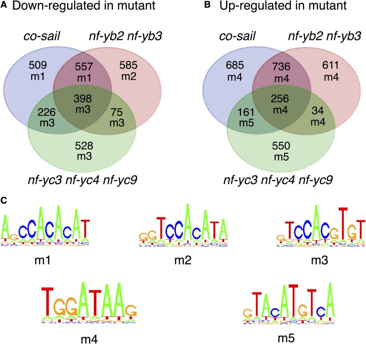
Nuclear Factor Y (NF-Y) is a heterotrimeric transcription factor that binds CCAAT elements. The NF-Y trimer is composed of a Histone Fold Domain (HFD) dimer (NF-YB/NF-YC) and NF-YA, which confers DNA sequence specificity. NF-YA shares a conserved domain with the CONSTANS, CONSTANS-LIKE, TOC1 (CCT) proteins. We show that CONSTANS (CO/B-BOX PROTEIN1 BBX1), a master flowering regulator, forms a trimer with Arabidopsis thaliana NF-YB2/NF-YC3 to efficiently bind the CORE element of the FLOWERING LOCUS T promoter. We term this complex NF-CO. Using saturation mutagenesis, electrophoretic mobility shift assays, and RNA-sequencing profiling of co, nf-yb, and nf-yc mutants, we identify CCACA elements as the core NF-CO binding site. CO physically interacts with the same HFD surface required for NF-YA association, as determined by mutations in NF-YB2 and NF-YC9, and tested in vitro and in vivo. The co-7 mutation in the CCT domain, corresponding to an NF-YA arginine directly involved in CCAAT recognition, abolishes NF-CO binding to DNA. In summary, a unifying molecular mechanism of CO function relates it to the NF-YA paradigm, as part of a trimeric complex imparting sequence specificity to HFD/DNA interactions. It is likely that members of the large CCT family participate in similar complexes with At-NF-YB and At-NF-YC, broadening HFD combinatorial possibilities in terms of trimerization, DNA binding specificities, and transcriptional regulation.


Figures

Figure 1.
Figure 1.
CO Binds DNA as a Trimer with At-NF-YB2/NF-YC3 and Recognizes the CORE Element. (A) CO forms a trimer with At-NF-YB2/NF-YC3 HFD binding to the FT CORE2 element. EMSAs were performed using fluorescently labeled FT CORE2 (lanes 1–14) or FT CCAAT (lanes 15–28) 31-mer oligonucleotide DNA probes (20 nM) by addition of the indicated proteins. CO-CCT (CO) was incubated at increasing concentrations (90, 180, 270, and 360 nM) with the CORE2 probe in the absence (lanes 2–5) or presence (lanes 9–12) of the At-NF-YB2/NF-YC3 HFD dimer (At-NF-YB2/YC3, 60 nM). As controls, At-NF-YA2 or -YA6 (YA2, YA6) was incubated with the CORE2 probe at the highest concentration of the dose curve (360 nM), with or without (−) the HFD dimer (60 nM) (YA2: lanes 13, 6; YA6: lanes 14, 7, respectively). Lane 1: CORE2 probe alone, without protein additions. DNA binding of CO or At-NF-YAs, as indicated (lanes 16–27), was assayed on the FT CCAAT probe in the presence of At-NF-YB2/NF-YC3 (60 nM), with the same protein concentration dose curve (90, 180, 270, and 360 nM). As controls, the FT CCAAT probe was incubated with the HFD dimer alone (60 nM, lane 15) or with At-NF-YA2 protein (360 nM, lane 28). NF-CO and NF-Y/DNA complexes are indicated by closed or open arrowheads. fp, free probe. (B) EMSA competition analysis of the At-NF-YB2/NF-YC3/CO complex specificity on the labeled CORE2 probe. Top panel: Sequences of the 31-mer CORE2 probe and unlabeled competitor derived from the FT promoter (−172/−141 from ATG). Oligos 1 to 6: CORE2 30-mers and 25-mer, the wild type or mutant was used as unlabeled competitors. The 31-mer derived from the FT enhancer CCAAT sequence and the FT mutant competitor (Cao et al., 2014) are listed below, together with the Hsp70 CCAAT competitor. Sequence identity with the probe is indicated by dots, and 5′ or 3′ sequence extensions, or mutated nucleotides are indicated in capital letters. The previously described TTGTGGTT CORE element (Tiwari et al., 2010) and the CCAAT pentamer are highlighted in bold letters. Bottom panel: EMSA competition analysis was performed by incubation of the CORE2 probe with the trimer composed of indicated subunits (At-NF-YB3/NF-YC3, 60 nM; CO, 180 nM -At-NFY-B2/YC3/CO-: lanes 2–27) in the presence of TE buffer alone (lanes 2 and 27) or with the addition of increasing concentrations of the indicated unlabeled competitors (5× or 25× molar excess; lanes 3–26). Lanes 1 and 28: CORE2 probe alone, without protein addition. The NF-CO/DNA complex is indicated by an arrowhead.
Figure 2.
Figure 2.
Determination of NF-CO Sequence Specificity in Vitro. (A) CORE2 competitors and mutagenesis strategy. The unlabeled wild-type CORE2 and 30-mer oligo sequences with mutated nucleotides are shown, as in Figure 1, for the three sets of CORE2 mutant oligos (mI, mII, and mIII). In the bar graphs, mI and mII mutant oligo competitor efficiency (competition) is expressed as ratio of the dose-response curve slope of the mutant versus the wild-type oligo (see Methods). Competition of the wild-type oligo is set as 1. Indicated values represent the mean of three (mI oligos; top panel) or two (mII oligos; bottom panel) series of competition assay experiments (see also Supplemental Figure 4). Error bars indicate ± sd for mI oligos or value ranges for mII oligos. Sequences in red boxes highlight mutations with reduced competition (<0.67 of wild-type oligo efficiency). (B) and (C) CORE2 mIII mutant oligo EMSA competition results. Competition efficiencies are shown as mean value of three independent series of experiments for each of the mIII single nucleotide mutant oligo, as indicated for (A). For each mutated position, the wild-type oligonucleotide competition value, set as 1, is also shown. Values are also displayed in the bar graph in (C) (average of three independent sets of experiments ± sd). For each nucleotide position, dark and light shaded bars denote mutant and wild-type (asterisk) competitors, respectively (see also Supplemental Figure 4). In (C), the sequence matrix obtained with the mIII competitions (information content) is shown on the right, for the sense (+) and reverse (−) strands of the FT promoter.
Figure 3.
Figure 3.
Identification of CO and HFD Matrices by RNA-Seq Analysis. RNA-seq analysis of differentially expressed genes in the co-sail, nf-yb2 nf-yb3, and nf-yc3 nf-yc4 nf-yc9 lines compared with wild-type Arabidopsis. The motifs enriched in the associated promoters are shown for each intersection of coregulated genes. (A) and (B) Venn diagram showing numbers of differentially expressed genes identified in comparisons between tested lines and wild-type plants, and overlaps between differentially expressed gene sets. For each gene set, an alphanumeric code signifies the most highly enriched motif identified. (C) Sequence logos describing the motifs identified from analyses of promoters (from −1000 to TSS/ATG) of DE gene sets (Supplemental Figure 6).
Figure 4.
Figure 4.
CO Binds the FT Promoter in an At-NF-YB2-NF-YB3-Dependent Manner. ChIP was performed on Col-0 (parental) and nf-yb2 nf-yb3 plants transgenic for p35S:CO-YFP/HA. Enrichment of the selected segments -CCAAT/-5.3kb, core promoter, exon 4/+2.0kb- were evaluated by qPCR with appropriate amplicons. Error bars indicate se with five biological replicates. In each replicate, three technical replicates were performed. Statistical significance was obtained using Bio-Rad CFX Manager Version 3.0; in each case, the comparison is between the nonimmune control (NIC) and the immunoprecipitation (IP). ***P < 0.001.
Figure 5.
Figure 5.
Properties of At-NF-YC9 Trimerization Mutants. (A) Alignment of At-NF-YCs. Multiple sequence alignment of NF-YC protein core domains. Multiple sequence alignment was computed using ClustalW in Geneious version 7.0. Amino acid residue positions of the HFD are indicated for the At-NF-YC9 and human proteins. Amino acids making physical contact with NF-YA are annotated by an asterisk (Nardini et al., 2013). Arrows mark the position of mutated residues, with the closed arrow indicating the conserved phenylalanine required for interaction with NF-YA in mammals, which was mutated in NF-YC9F151R V153K and in the recombinant At-NF-YC9F151R HFD protein. At, Arabidopsis thaliana; Hs, Homo sapiens. (B) In vivo analysis of timing of flowering. T1 generation flowering time analysis of pNF-YC9:NF-YC9F151R V153K in the nf-yc triple (nf-yc3 nf-yc4 nf-yc9) mutant background. Asterisks represent significant differences derived from one-way ANOVA (P < 0.05) followed by Dunnett’s multiple comparison post hoc tests against the nf-yc triple mutant. (C) Expression of At-NF-YC9 transgenes in transgenic plants. Protein expression in the plant lines used for the flowering time analysis was analyzed by immunoblot with antibodies directed to a translationally fused HA-epitope (top panel). Protein loading and transfer was assessed by Ponceau staining (bottom panel). (D) Y2H assays of At-NF-YC9. Full-length NF-YC9 and mutant variants tested using Y2H against empty vector (EV) control, NF-YB2, NF-YA1, NF-YA2, and CO. Note that NF-YC9 has slight autoactivation. (E) EMSAs on CORE2 and CCAAT of wild-type and mutant At-NF-YC9. Trimerization and DNA binding of the At-NF-YC9F151R mutant (YC9F151R; lanes 7–11) or the wild type (YC9; lanes 2–6) containing HFD dimer (60 nM) was assessed the with the CORE2 probe (lanes 1–14), by addition of the CO subunit at increasing concentrations (90, 180, 270, and 360 nM; lanes 3–6 and 8–11). At-NF-YA2 (YA2) trimerization with the wild-type or mutant dimers (lanes 17–20 and 22–25, respectively) was assessed with the CCAAT probe (lanes 15–28). As negative controls, CO or At-NF-YA2 was added alone to the reaction with the respective probes (lanes 12 and 26). At-NF-YC9 trimer specificity was also assessed by addition of the CO or At-NF-YA2 containing trimers to the CCAAT or CORE2 probe, respectively (lanes 28 and 14). DNA binding of At-NF-YC3 (YC3) containing trimers was also used as internal control (lanes 13 and 27). In lanes 2, 7, 16, and 21, wild-type or mutant HFD dimers were incubated alone with the probe. NF-CO and NF-Y/DNA complexes are indicated by labeled arrowheads. Lanes 1 and 15: CORE2 and CCAAT probes without protein additions. fp, free probe.
Figure 6.
Figure 6.
Properties of At-NF-YB2 Trimerization Mutant. Trimerization and DNA binding of the E65R mutant (YB2E65R; lanes 7–11) or wild-type (lanes 2–6) At-NF-YB2 containing HFD dimer (60 nM) was assessed with the CORE2 probe (lanes 1–14), by addition of the CO subunit at increasing concentrations (90, 180, 270, and 360 nM; lanes 3–6 and 8–11). At-NF-YA2 (YA2) trimerization with the wild-type or mutant dimers (lanes 17–20 and 22–25, respectively) was assessed with the CCAAT probe (lanes 15–28). As negative controls, CO or At-NF-YA2 was added alone to the reaction with the respective probes (lanes 12 and 26). Trimer specificity was assessed by addition of the CO or At-NF-YA2 containing trimers to the CCAAT or CORE2 probe, respectively (lanes 13, 14, 27, and 28 as indicated). In lanes 2, 7, 16, and 21, wild-type or mutant HFD dimers were incubated alone with the probe. NF-CO and NF-Y/DNA complexes are indicated by labeled arrowheads. Lanes 1 and 15: CORE2 and CCAAT probes without protein additions. fp, free probe.
Figure 7.
Figure 7.
The CO CCT Drives Sequence Specificity of NF-CO. (A) CO mutation R340Q of co-7 abolishes NF-CO DNA binding. Wild-type or R340Q CO was incubated at increasing concentrations (90, 180, 270, and 360 nM) with the CORE2 probe in the absence (−) (lanes 2–9) or presence (lanes 11–18) of the At-NF-YB2/NF-YC3 HFD dimer (60 nM). In lane 10, the At-NF-YB2/NF-YC3 HFD dimer was incubated alone with the probe. Lane 1: probe alone, without protein additions. fp, free probe. (B) Schematic representation of selected NF-YA interactions with the C2A3 bp of CCAAT. Highlight of NF-YA A2 helix within the NF-Y/DNA 3D structure (PDB: 4AWL), with interactions of Arg-281, Arg-283 (corresponding to CO Arg-340), and Gly-287 (CO Gly-343) amino acid residues (indicated in single letter code: Arg-281, Arg-283, and Gly-287 respectively) with the G2 and A3 nucleotides. NF-YA protein (cyan) and the sugar-phosphate DNA strand backbones are represented as colored strings, with the CCAAT (I) and complementary (J) strands in red and green, respectively. Orientation of DNA strands is indicated. Selected NF-YA residues and nitrogen bases of the C2A3:G2T3 nucleotides are labeled and displayed in ball and stick model in color matching the main chain color code. Gly-287 main chain and Arg-283 side chain contacts with G2 atoms, and Arg-281 side chain with A3, are indicated by gray lines (Nardini et al., 2013). The NF-YB/NF-YC subunits within the 4AWL structure were omitted for clarity. The image was obtained with Protein Workshop (Moreland et al., 2005). (C) Amino acid sequence alignment of the C-terminal portion of CO CCT domain with mammalian NF-YA homology region is shown, with the proposed sequence-specific interactions, based on the NF-Y/DNA complex crystal structure (PDB: 4AWL). DNA sequence of the NF-CO and NF-Y respective element is shown at the top and bottom of the alignment, with the indicated orientation of the DNA strands and base-pair positions in the bound elements numbered (see text). Side chain interactions of NF-YA with the CCAAT bases are indicated by full lines (bottom). On top of the alignment, dashed lines represent potential CO residues interactions with the CORE matrix. Conserved and nonconserved residues are highlighted in green and blue, respectively. R340Q in co-7 is indicated on top of the alignment. The closed circle represents hydrophobic base stacking interactions of phenylalanine residue side chains with the CA:GT nucleotides. Bold nucleotides highlight the divergence in sequence specificity of the two complexes.
Figure 8.
Figure 8.
Scheme of NF-Y versus NF-CO Specificity. Association of CO or NF-YA with NF-YB/NF-YC dimers provides robust and specific recognition of the respective DNA element by the trimeric NF-CO and NF-Y complexes.

Comment in

Similar articles

Cited by

References

    1. Abelenda J.A., Cruz-Oró E., Franco-Zorrilla J.M., Prat S. (2016). Potato StCONSTANS-like1 suppresses storage organ formation by directly activating the FT-like StSP5G repressor. Curr. Biol. 26: 872–881. - PubMed
    1. Adrian J., Farrona S., Reimer J.J., Albani M.C., Coupland G., Turck F. (2010). cis-Regulatory elements and chromatin state coordinately control temporal and spatial expression of FLOWERING LOCUS T in Arabidopsis. Plant Cell 22: 1425–1440. - PMC - PubMed
    1. Ben-Naim O., Eshed R., Parnis A., Teper-Bamnolker P., Shalit A., Coupland G., Samach A., Lifschitz E. (2006). The CCAAT binding factor can mediate interactions between CONSTANS-like proteins and DNA. Plant J. 46: 462–476. - PubMed
    1. Blackman B.K., Michaels S.D. (2010). Does CONSTANS act as a transcription factor or as a co-activator? The answer may be--yes. New Phytol. 187: 1–3. - PubMed
    1. Bu Z., Yu Y., Li Z., Liu Y., Jiang W., Huang Y., Dong A.W. (2014). Regulation of arabidopsis flowering by the histone mark readers MRG1/2 via interaction with CONSTANS to modulate FT expression. PLoS Genet. 10: e1004617. - PMC - PubMed

MeSH terms