. 2017 Jul 7;292(27):11547-11560.
doi: 10.1074/jbc.M116.774067. Epub 2017 May 19.

Functional regions of the peroxin Pex19 necessary for peroxisome biogenesis

Affiliations

Functional regions of the peroxin Pex19 necessary for peroxisome biogenesis

Gaurav Agrawal et al. J Biol Chem. .

Abstract

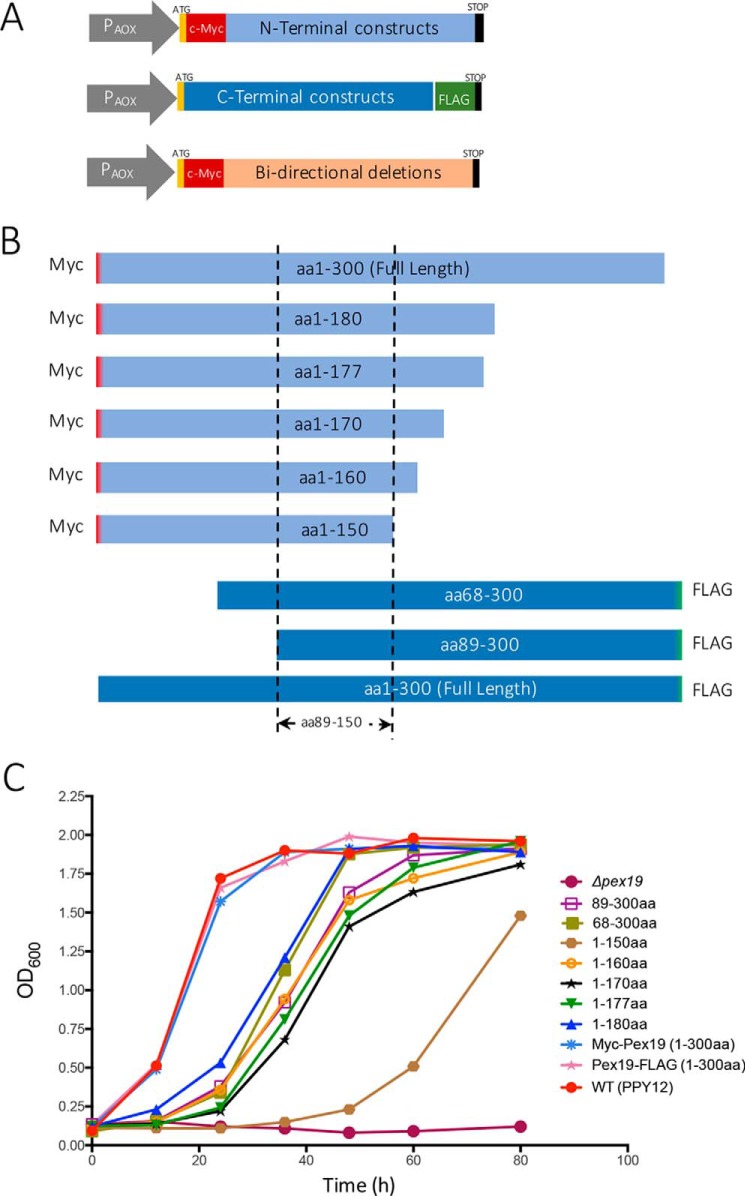
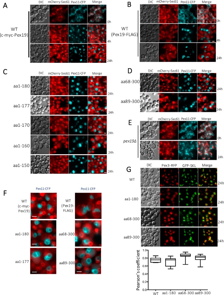
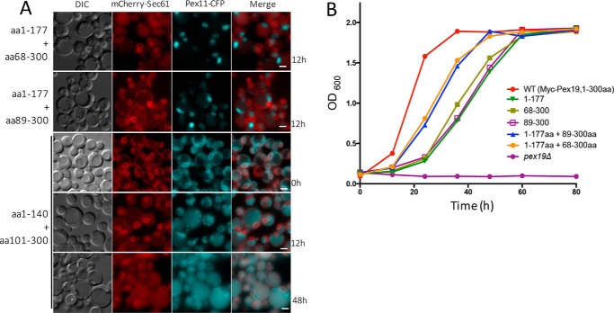
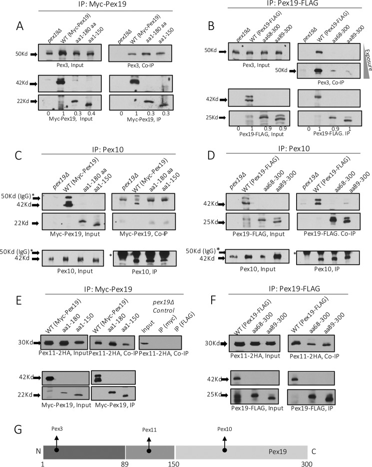
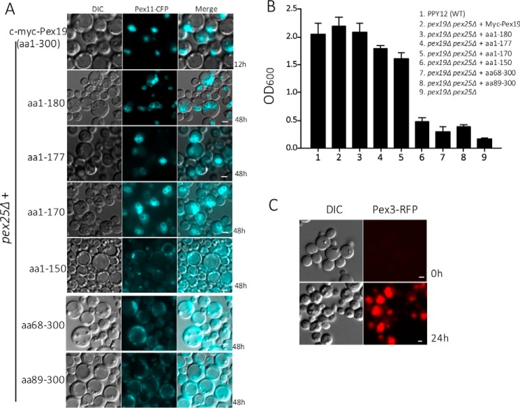
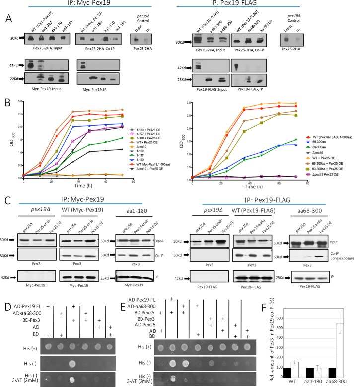
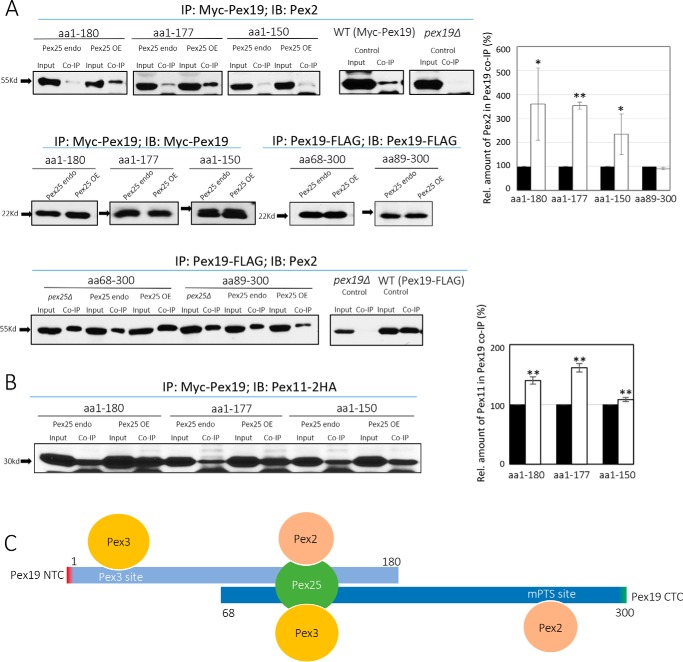
The peroxins Pex19 and Pex3 play an indispensable role in peroxisomal membrane protein (PMP) biogenesis, peroxisome division, and inheritance. Pex19 plays multiple roles in these processes, but how these functions relate to the structural organization of the Pex19 domains is unresolved. To this end, using deletion mutants, we mapped the Pex19 regions required for peroxisome biogenesis in the yeast Pichia pastoris Surprisingly, import-competent peroxisomes still formed when Pex19 domains previously believed to be required for biogenesis were deleted, although the peroxisome size was larger than that in wild-type cells. Moreover, these mutants exhibited a delay of 14-24 h in peroxisome biogenesis. The shortest functional N-terminal (NTCs) and C-terminal constructs (CTCs) were Pex19 (aa 1-150) and Pex19 (aa 89-300), respectively. Deletions of the N-terminal Pex3-binding site disrupted the direct interactions of Pex19 with Pex3, but preserved interactions with a membrane peroxisomal targeting signal (mPTS)-containing PMP, Pex10. In contrast, deletion of the C-terminal mPTS-binding domain of Pex19 disrupted its interaction with Pex10 while leaving the Pex19-Pex3 interactions intact. However, Pex11 and Pex25 retained their interactions with both N- and C-terminal deletion mutants. NTC-CTC co-expression improved growth and reversed the larger-than-normal peroxisome size observed with the single deletions. Pex25 was critical for peroxisome formation with the CTC variants, and its overexpression enhanced their interactions with Pex3 and aided the growth of both NTC and CTC Pex19 variants. In conclusion, physical segregation of the Pex3- and PMP-binding domains of Pex19 has provided novel insights into the modular architecture of Pex19. We define the minimum region of Pex19 required for peroxisome biogenesis and a unique role for Pex25 in this process.

Keywords: membrane biogenesis; organelle; peroxisome; protein assembly; trafficking.


Conflict of interest statement

The authors declare that they have no conflicts of interest with the contents of this article

Figures

Figure 1.
Figure 1.
Schematic representations of Pex19 deletion constructs. A, all the deletion constructs were expressed from the inducible alcohol oxidase promoter (PAOX) in pex19Δ cells. The NTCs and the bidirectional deletion constructs each had an N-terminal c-myc tag, whereas the CTCs each had a C-terminal FLAG tag. All the constructs had a start (ATG) and a stop codon (TAA), at the beginning and end of the ORF, respectively. B, schematic representation of the functional Pex19 deletion constructs. All the functional deletion constructs have an overlap between aa 89 and 150. C, the pex19Δ cells transformed with plasmids expressing specified deletion constructs from chromosomally-integrated plasmid DNA, were assayed for growth on methanol medium. Cells were grown overnight in YPD and ∼0.1 A600/ml was further inoculated into methanol medium. Cell growth was measured at specific times. The experiment was repeated three times with similar results. P. pastoris PPY12 cells were used as WT control.
Figure 2.
Figure 2.
Fluorescence microscopy analysis of methanol-grown cells expressing the specified fluorescently-tagged proteins. All the constructs were expressed in the pex19Δ cells. Cells were grown in YPD and switched during exponential phase to methanol medium. DIC, differential interference contrast. Scale bar, 2 μm. A and B, localization of Pex11-CFP and Sec61-mCherry, the ER marker, in pex19Δ cells expressing full-length Pex19 constructs at specified times. Pex11-CFP colocalized with the ER marker at 0 h only in the absence of Pex19, but it did not colocalize at 4 or 24 h as it was peroxisomal. C and D, absence of co-localization of Pex11-CFP with Sec61-mCherry in NTC and CTC variants capable of growing on methanol. Pex11-CFP is peroxisomal in these strains because the Pex19 constructs retain their ability to facilitate peroxisome biogenesis. E, co-localization of Pex11-CFP and mCherry-Sec61 in methanol-grown pex19Δ cells after 24 h. F, larger than normal peroxisomes labeled with Pex11-CFP, relative to those in WT cells, in NTC and CTC variants competent to form peroxisomes. G, evidence of bona fide peroxisome formation in WT and specified NTC and CTC variants of Pex19, as judged by the colocalization of peroxisomal membrane marker, Pex3-RFP, and the peroxisomal matrix marker, GFP-SKL. Also shown is colocalization of Pex3-RFP and GFP-SKL in Pex19 variants. Pearson's correlation coefficients were calculated using the coloc2 plug-in for ImageJ, and the data are displayed as interquartile boxes and whisker plots. Pex3-RFP was expressed from the PAOX promoter, and GFP-SKL was expressed from the PGAP promoter.
Figure 3.
Figure 3.
Fluorescence microscopy and growth analysis of P. pastoris strains co-expressing the specified NTC and CTC variants in pex19Δ cells. Cells were grown in YPD and switched during exponential phase to methanol medium. A, fluorescence microscopy analysis of methanol-grown cells co-expressing the specified NTC and CTC variants. Pex11-CFP and Sec61-mCherry co-localized only at 0 h, when Pex19 was absent, but this co-location was lost as peroxisomes formed and Pex11-CFP became peroxisomal. Scale bar: 2 μm. B, cells co-expressing the specified NTC and CTC variants were assayed for growth on methanol medium as described in the legend to Fig. 1B. The experiment was repeated three times with similar results.
Figure 4.
Figure 4.
Interaction of WT Pex19 and its NTC and CTC variants with different PMPs. All the constructs were expressed in pex19Δ cells. Co-immunoprecipitations were performed with appropriate antibodies against c-myc-Pex19 (A and E), Pex19-FLAG (B and F), or Pex10 (C and D) as described under ”Materials and methods.“ The immunoprecipitates were then immunoblotted with appropriate antibodies to detect Pex3 (A and B), Myc-Pex19 (C), Pex19-FLAG (D), and Pex11–2HA (E and F). The proteins detected in the Input, IP, and co-IP are shown. The relative protein abundances of WT Pex19 and its variants, as detected by quantitation of the protein band intensities, are shown by numbers in A and B. The WT Pex19 level was set to 1 and Pex19 in pex19Δ cells to 0. The co-IP was repeated three times with similar qualitative results regarding the presence or absence of the proteins that co-immunoprecipitated with Pex19 or its variants. G, schematic representation of the binding sites on Pex19 for different PMPs.
Figure 5.
Figure 5.
Pex25 is required for peroxisome biogenesis in the CTC variants of Pex19. A, localization of Pex11-CFP in methanol-grown pex19Δ pex25Δ cells expressing either the WT Pex19 or the specified NTCs or CTCs (scale bar: 2 μm). Note that only one NTC (aa 1–150) and two CTC variants (aa 68–300 and aa 89–300 of Pex19) expressing Pex11-CFP did not show punctate, peroxisome-like structures. B, methanol-grown pex19Δ pex25Δ cells expressing constructs described in A were assayed for growth in methanol. Note that only those Pex19 variants that failed to localize Pex11-CFP to punctate peroxisomes in A failed to grow in methanol. The experiment was repeated three times with similar results. C, localization of Pex3-RFP, expressed from the inducible PAOX promoter, to punctate, peroxisome-like structures in methanol-grown pex3Δ pex25Δ cells. Cells were grown in YPD and switched during exponential phase to methanol medium.
Figure 6.
Figure 6.
Pex25 is required for restoring the interaction between CTC variants and Pex3. A, co-immunoprecipitation was performed by immunoprecipitating either Myc-Pex19 (for NTCs, left panel) or Pex19-FLAG (for the CTCs, right panel) using appropriate antibodies, as described under ”Materials and methods.“ Immunoblotted proteins detected in the Input, IP, and co-IP samples are shown. The co-IP was repeated three times with similar results. B, methanol-grown pex19Δ cells, either expressing Pex25 from the constitutive GAP promoter (Pex25 OE) or from the endogenous (endo) promoter, with the WT Pex19 or the specified NTCs or the CTCs were assayed for growth in methanol medium. The experiment was repeated three times with similar results. C, co-immunoprecipitation was performed by immunoprecipitating either Myc-Pex19 (for NTCs, left panel) or Pex19-FLAG (for the CTCs, right panel) in strains expressing different Pex25 levels. Immunoblots were then done to detect Pex3 levels in the co-IP. The co-IP was repeated three times with similar results. D and E, yeast two-hybrid assay of Pex19 full-length (FL), Pex19(68–300), Pex3, and Pex25. F, effect of Pex25 overexpression on Pex3-Pex19 co-immunoprecipitation. The relative abundance of Pex3 in the Pex19 co-immunoprecipitates in strains expressing either endogenous (black bars) or overexpressed (white bars) levels of Pex25 for Pex19 variants depicted in C was quantitated. The Pex3 levels of cells expressing endogenous levels of Pex25 were set to 100%. Error bars represent mean ± S.E. of three independent experiments.
Figure 7.
Figure 7.
Effect of Pex25 on the ability of Pex19 variants to bind other PMPs. A, co-immunoprecipitation of Myc-Pex19 or Pex19-FLAG with Pex2, in strains expressing different levels of Pex25. The bar graph depicts the levels of Pex2 in the co-IP of Pex19 variants, in the presence of either endogenous (black bars) or overexpressed (OE) levels (white bars) of Pex25. *, p < 0.05; **, p < 0.01. B, co-immunoprecipitation of Myc-Pex19 NTCs with Pex11-2HA, in strains expressing different levels of Pex25. The bar graph depicts the levels of Pex11-2HA in the co-IP of Pex19 variants, in the presence of either endogenous (black bars) or overexpressed levels (white bars) of Pex25. *, p < 0.05; **, p < 0.01. The co-immunoprecipitations were repeated twice with similar results. C, schematic representation of key binding sites missing in the Pex19 NTC and CTC variants. Pex25 binding to the central domain of Pex19 bridges the missing PMP (e.g. Pex2) and Pex3 interactions in the NTC and CTC, respectively, and explains why Pex25 overexpression stimulates growth of cells expressing these constructs, relative to strains expressing endogenous levels of Pex25. The direct binding of Pex3 and Pex2/Pex10 to Pex19 through the sites shown is the preferred mode of binding, because pex25Δ cells show no defect in peroxisome biogenesis, but the bridging interactions mediated by Pex25 become prominent only when the Pex3 site and/or the PMP site in Pex19 is missing.

References

    1. Subramani S. (1998) Components involved in peroxisome import, biogenesis, proliferation, turnover, and movement. Physiol. Rev. 78, 171–188 - PubMed
    1. Wanders R. J., and Waterham H. R. (2006) Biochemistry of mammalian peroxisomes revisited. Annu. Rev. Biochem. 75, 295–332 - PubMed
    1. Schrader M., and Fahimi H. D. (2008) The peroxisome: still a mysterious organelle. Histochem. Cell Biol. 129, 421–440 - PMC - PubMed
    1. Klouwer F. C., Berendse K., Ferdinandusse S., Wanders R. J., Engelen M., and Poll-The B. T. (2015) Zellweger spectrum disorders: clinical overview and management approach. Orphanet J. Rare Dis. 10, 151. - PMC - PubMed
    1. Schrader M., and Fahimi H. D. (2006) Growth and division of peroxisomes. Int. Rev. Cytol. 255, 237–290 - PubMed

Publication types

LinkOut - more resources