The origin and evolution of human glutaminases and their atypical C-terminal ankyrin repeats
- PMID: 28526749
- PMCID: PMC5500818
- DOI: 10.1074/jbc.M117.787291
The origin and evolution of human glutaminases and their atypical C-terminal ankyrin repeats
Abstract
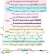
On the basis of tissue-specific enzyme activity and inhibition by catalytic products, Hans Krebs first demonstrated the existence of multiple glutaminases in mammals. Currently, two human genes are known to encode at least four glutaminase isoforms. However, the phylogeny of these medically relevant enzymes remains unclear, prompting us to investigate their origin and evolution. Using prokaryotic and eukaryotic glutaminase sequences, we built a phylogenetic tree whose topology suggested that the multidomain architecture was inherited from bacterial ancestors, probably simultaneously with the hosting of the proto-mitochondrion endosymbiont. We propose an evolutionary model wherein the appearance of the most active enzyme isoform, glutaminase C (GAC), which is expressed in many cancers, was a late retrotransposition event that occurred in fishes from the Chondrichthyes class. The ankyrin (ANK) repeats in the glutaminases were acquired early in their evolution. To obtain information on ANK folding, we solved two high-resolution structures of the ANK repeat-containing C termini of both kidney-type glutaminase (KGA) and GLS2 isoforms (glutaminase B and liver-type glutaminase). We found that the glutaminase ANK repeats form unique intramolecular contacts through two highly conserved motifs; curiously, this arrangement occludes a region usually involved in ANK-mediated protein-protein interactions. We also solved the crystal structure of full-length KGA and present a small-angle X-ray scattering model for full-length GLS2. These structures explain these proteins' compromised ability to assemble into catalytically active supra-tetrameric filaments, as previously shown for GAC. Collectively, these results provide information about glutaminases that may aid in the design of isoform-specific glutaminase inhibitors.
Keywords: X-ray crystallography; cancer; glutaminase; human; isoform; metabolism.
© 2017 by The American Society for Biochemistry and Molecular Biology, Inc.
Conflict of interest statement
The authors declare that they have no conflicts of interest with the contents of this article
Figures


References
-
- Bergström J., Fürst P., Norée L. O., and Vinnars E. (1974) Intracellular free amino acid concentration in human muscle tissue. J. Appl. Physiol. 36, 693–697 - PubMed
-
- Levine A. J., and Puzio-Kuter A. M. (2010) The control of the metabolic switch in cancers by oncogenes and tumor suppressor genes. Science 330, 1340–1344 - PubMed
-
- Wise D. R., DeBerardinis R. J., Mancuso A., Sayed N., Zhang X.-Y., Pfeiffer H. K., Nissim I., Daikhin E., Yudkoff M., McMahon S. B., and Thompson C. B. (2008) Myc regulates a transcriptional program that stimulates mitochondrial glutaminolysis and leads to glutamine addiction. Proc. Natl. Acad. Sci. U.S.A. 105, 18782–18787 - PMC - PubMed
Publication types
MeSH terms
Substances
Associated data
- Actions
- Actions
- Actions
- Actions
- Actions
- Actions
- Actions
- Actions
- Actions
Grants and funding
LinkOut - more resources
Full Text Sources
Other Literature Sources
Molecular Biology Databases

