. 2017 May 19;7(1):2149.
doi: 10.1038/s41598-017-02202-4.

EndophilinAs regulate endosomal sorting of BDNF-TrkB to mediate survival signaling in hippocampal neurons

Affiliations

EndophilinAs regulate endosomal sorting of BDNF-TrkB to mediate survival signaling in hippocampal neurons

Katja Burk et al. Sci Rep. .

Abstract

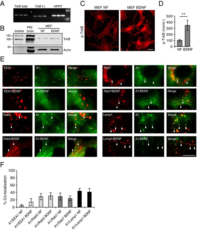
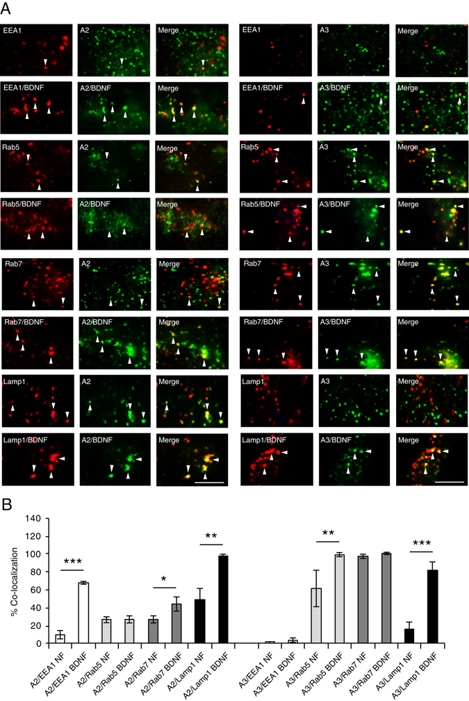
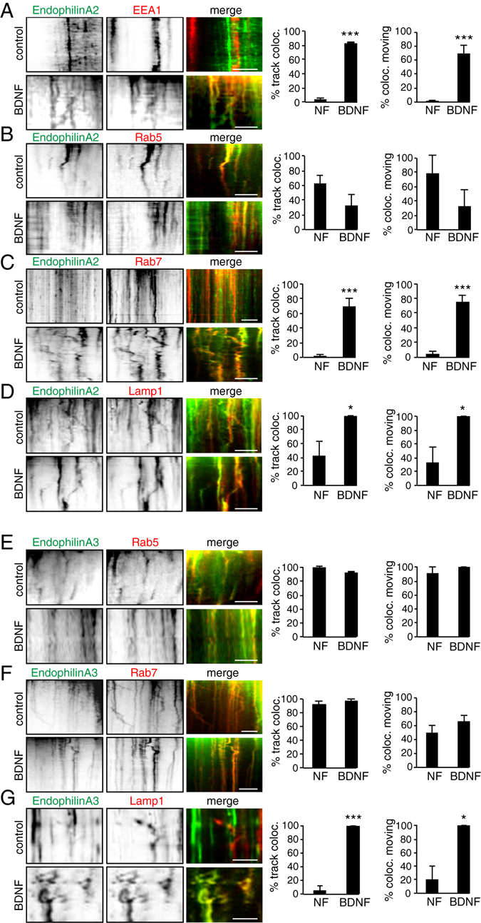
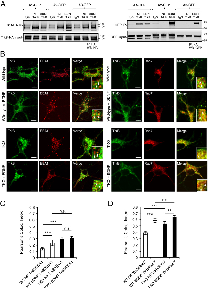
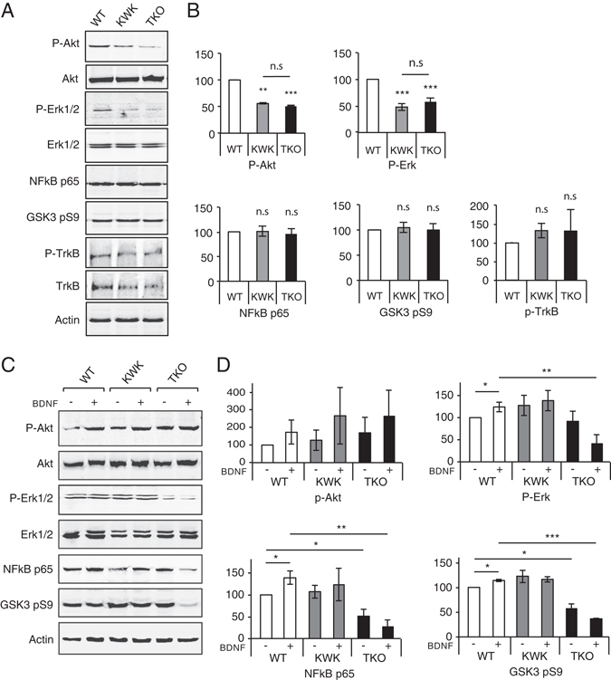
The sorting of activated receptors into distinct endosomal compartments is essential to activate specific signaling cascades and cellular events including growth and survival. However, the proteins involved in this sorting are not well understood. We discovered a novel role of EndophilinAs in sorting of activated BDNF-TrkB receptors into late endosomal compartments. Mice lacking all three EndophilinAs accumulate Rab7-positive late endosomes. Moreover, EndophilinAs are differentially localized to, co-traffic with, and tubulate, distinct endosomal compartments: In response to BDNF, EndophilinA2 is recruited to both early and late endosomes, EndophilinA3 is recruited to Lamp1-positive late endosomes, and co-trafficks with Rab5 and Rab7 in both the presence and absence of BDNF, while EndophilinA1 colocalizes at lower levels with endosomes. The absence of all three EndophilinAs caused TrkB to accumulate in EEA1 and Rab7-positive endosomes, and impaired BDNF-TrkB-dependent survival signaling cascades. In addition, EndophilinA triple knockout neurons exhibited increased cell death which could not be rescued by exogenous BDNF, in a neurotrophin-dependent survival assay. Thus, EndophilinAs differentially regulate activated receptor sorting via distinct endosomal compartments to promote BDNF-dependent cell survival.


Conflict of interest statement

The authors declare that they have no competing interests.

Figures

Figure 1
Figure 1
Rab7-positive endosomes accumulate in EndophilinA TKO hippocampal neurons. (A) Identification of endosomal structures in wild-type and EndophilinA TKO neurons using immunocytochemistry to detect endogenous levels of the endosomal markers EEA1, and Rab7. Representative images of WT and Endophilin TKO hippocampal neurons stained for EEA1 and Rab7 are shown. Map2 antibody was used to label neurons. Scale bar = 10 μm; (B) Quantification of endosomal marker signal for EEA1 or Rab7 (C) normalized to area. Significance determined by unpaired two-tailed Student’s t-test comparing each condition to control, n = 30 images per condition from 3 independent cell cultures; error = SEM, **p < 0.01. (C) Western blots showing levels of EEA1 and Rab7 in wild-type and EndophilinA TKO brain homogenates. (D) Quantitation of protein levels of EEA1 and Rab7 (F) in control and EndophilinA TKOs normalized to actin; n = 3 independent brain samples, error = SEM, *p < 0.05, **p < 0.01.
Figure 2
Figure 2
EndophilinA1 colocalizes with endosomes. (A) qPCR products of total TrkB (including full length and truncated isoforms), full length TrkB (TrkB f.l.) and HPRT as a control from mouse embronic fibroblast (MEF) lysates. (B) Western blot of TrkB in MEF lysates in control no factor (NF) conditions, and following treatment with BDNF. P60 brain lysate serves as a positive control, and actin as a loading control. (C) Images of pTrkB immunostaining in cultured MEFs in “no factor” (NF) untreated conditions, or following treatment with 100 ng/ml BDNF for 30 min; scale bar = 10 µm. (D) Quantitation of pTrkB signal in MEFs in NF and BDNF conditions; n-21 images per condition, error = SEM, significance determined by Student’s t-test. (E) Representative TIRF microscopy images of MEFs co-transfected with GFP-tagged EndophilinA1 and the RFP-tagged endosomal markers EEA1, Rab5, Rab7 or Lamp1, in the presence and absence of 100 ng/ml BDNF. Arrows indicate colocalization of EndophilinA1 (green) with endosomal markers (red). (F) Quantitation of colocalization of transfected GFP-tagged EndophilinA1 with RFP-tagged EEA1, Rab5, Rab7, or Lamp1 in control conditions with no factors (NF) or following addition of BDNF; n = 8–10 images per condition from 3 independent cell cultures, significance determined by unpaired two-tailed Student’s t-test comparing non-stimulated to stimulated conditions, error = SEM, scale bar = 10 µm.
Figure 3
Figure 3
EndophilinA2 and A3 are recruited to endosomes in response to BDNF. (A) Representative TIRF microscopy images of MEFs co-transfected with GFP-tagged EndophilinA2 (left panels) or EndophilinA3 (right panels) and the RFP-tagged endosomal markers EEA1, Rab5, Rab7 or Lamp1, in the presence and absence of 100 ng/ml BDNF. Arrows indicate colocalization of EndophilinA (green) with endosomal markers (red). (B) Quantitation of colocalization of transfected GFP-tagged EndophilinA2, and A3 with RFP-tagged EEA1, Rab5, Rab7 and Lamp1; n = 8–10 images per condition from 3 independent cell cultures, significance determined by unpaired two-tailed Student’s t-test test comparing non-stimulated to stimulated conditions, error = SEM, *p < 0.05, **p < 0.01, ***p < 0.001, scale bar = 10 µm.
Figure 4
Figure 4
EndophilinA2 and EndophilinA3 co-traffic with endosomes. (A) Kymographs (left panels) of co-trafficking of EndophilinA2 and endosomal markers before and after application of 100 ng/ml BDNF in 5 minute TIRF time-lapse images of MEFs co-transfected with GFP-tagged EndophilinA2 and RFP-tagged EEA1, Rab5 (B), Rab7 (C), or Lamp1 (D). Quantitation of recruitment of EndophilinA2 to endosomes and mobility of EndophilinA2-positive endosomes (before or after administering 100 ng/ml BDNF) by percent of EndophilinA2 and endosomal kymograph tracks colocalizing, and percent of co-localizing mobile endosomes (A–D, right panels). (E) Kymographs of co-trafficking of EndophilinA3 and RFP-tagged Rab5 (E), Rab7 (F), or Lamp1 (G). Quantitation of recruitment of EndophilinA3 to endosomes before and after administering BDNF, by percent of tracks colocalizing, and percent of co-localizing mobile endosomes (E–G, right panels); n = 8–10 images per condition from 3 independent cell cultures, significance determined by unpaired two-tailed Student’s t-test non-stimulated to stimulated conditions, error = SEM, *p < 0.05, **p < 0.01, ***p < 0.001, scale bar = 10 µm.
Figure 5
Figure 5
EndophilinAs tubulate endosomal compartments to which they are recruited. (A) Representative timecourse of a tubulation event of EndophilinA2 on a Rab7-positive endosome over 200 seconds in co-transfected EndophilinA TKO MEFs. Scale bar = 5 µm. (B) Example of tubulation events of EndophilinA1 on Lamp1-positive endosomes in the absence (NF) and presence of 100 ng/ml BDNF, of EndophilinA2 on Rab7 and Lamp1-positive endosomes in the absence (NF) and presence of BDNF (C), and of EndophilinA3 on EEA1, Rab7, and Lamp1-positive endosomes in the absence (NF) and presence of BDNF (D) Arrows indicate tubulation events in which endosomal markers and EndophilinA isoforms co-localize. Scale bars = 2 µm for (BD). (E) Quantitation of the percent increase in tubulation of endosomal compartments by EndophilinA1, EndophilinA2, and EndophilinA3, compared to EndophilinA triple knockouts in control no factor (NF) conditions and following 100 ng/ml BDNF treatment; n = 8–10 images per condition from 3 independent cell cultures, significance determined by paired two-tailed Student’s t-test, error = SEM, *p < 0.05, **p < 0.01, ***p < 0.001, scale bar = 10 µm.
Figure 6
Figure 6
TrkB receptors accumulate in EEA1 and Rab7 positive endosomes in EndophilinA triple knockouts. (A) HA-conjugated ProteinA beads (or IgG control beads) were used to pull down HA-TrkB (left) and EndophilinA1, A2, or A3-GFP (right) in co-transfected Hek293 cells in the presence or absence of 100 ng/ml BDNF. (B) Representative images of wild-type and EndophilinA TKO mouse neuronal cultures transfected with TrkB-GFP and the RFP-tagged endosomal markers EEA1 or Rab7. Arrows indicate TrkB colocalizing with EEA1 or Rab7. Scale bar = 10 µm. (C) Quantitation of colocalization of TrkB-GFP with RFP-tagged EEA1 or Rab7 (D), in wild-type and EndophilinA TKO neurons. In EndophilinA triple knockout neurons, more TrkB was found in EEA1 and Rab7 positive endosomes in both the presence and absence of BDNF compared to control; n = 30 images from 4 independent cultures, significance determined by unpaired two-tailed Student’s t-test compared to non-stimulated control and between non stimulated TKO and BDNF treated TKO, error = SEM; **p < 0.01, ***p < 0.001.
Figure 7
Figure 7
EndophilinA triple knockouts show changes in BDNF-mediated signaling. (A) Western blots of the indicated proteins in EndophilinA1/3 double knockout (KWK refers to Knockout, Wild-type, Knockout, for EndophilinA1, A2, A3), and EndophilinA triple knockout (TKO) brain lysates compared to wild-type. (B) Quantitation of P-Akt, P-Erk, NFkB p65, GSK3 pS9, and pTrkB protein levels in Western blots normalized to actin; n = 3 independent experiments, significance determined by unpaired two-tailed Student’s t-test comparing each condition to control, error = SEM, **p < 0.01, ***p < 0.001. (C) Western blots of the indicated proteins in EndophilinA1/3 double knockout (KWK refers to Knockout, Wild-type, Knockout, for EndophilinA1, A2, A3), and EndophilinA triple knockout (TKO) hippocampal cultures compared to wild-type in control and BDNF-treated conditions (100 ng/ml BDNF for 30 minutes). (D) Quantitation of P-Akt, P-Erk, NFkB p65 and GSK3 pS9 protein levels in Western blots normalized to actin; n = 3 independent experiments, significance determined by unpaired two-tailed Student’s t-test comparing each condition to control, error = SEM, **p < 0.01, ***p < 0.001.
Figure 8
Figure 8
Survival is altered in low-density cultures of EndophilinA triple knockout neurons. (A,B) Brain sections of wild-type and EndophilinA triple knockout mice immunostained with Neurotrace (green) and DAPI (red) showed no obvious defects in overall structure of the hippocampus or the number of Neurotrace/DAPI-positive cells (quantified in C,D). (E) High-density neuronal cultures of wild-type and EndophilinA triple knockout neurons immunostained for acetyl tubulin. Both wild-type and EndophilinA TKO neurons formed networks and survived up to 14 days in culture. (F) Survival assay of wild-type and Endophilin A1-/-; A2-/-; A3-/- low-density cultures: EndophilinA TKO neurons showed higher rates of cell death compared to wild-type neurons after 48 hours in culture in the absence of BDNF. This increase in cell death in Endophilin A1-/-; A2-/-; A3-/- neurons could not be rescued by administration of 100 ng/ml exogenous BDNF compared to control during a 120-hour time course; n = 4 independent experiments, significance determined by unpaired two-tailed Student’s t-test, comparing each condition to numbers of initially plated neurons. Error = SEM, *p < 0.05, **p < 0.01.

Similar articles

Cited by

References

    1. Andjelkovic M, et al. Nerve growth factor promotes activation of the alpha, beta and gamma isoforms of protein kinase B in PC12 pheochromocytoma cells. Eur. J Biochem. 1998;251:195–200. doi: 10.1046/j.1432-1327.1998.2510195.x. - DOI - PubMed
    1. Giachino C, et al. A novel SH3-containing human gene family preferentially expressed in the central nervous system. Genomics. 1997;41:427–434. doi: 10.1006/geno.1997.4645. - DOI - PubMed
    1. Ringstad N, Nemoto Y, De Camilli P. The SH3p4/Sh3p8/SH3p13 protein family: binding partners for synaptojanin and dynamin via a Grb2-like Src homology 3 domain. Proc. Natl. Acad. Sci. USA. 1997;94:8569–8574. doi: 10.1073/pnas.94.16.8569. - DOI - PMC - PubMed
    1. So CW, et al. Expression and protein-binding studies of the EEN gene family, new interacting partners for dynamin, synaptojanin and huntingtin proteins. Biochem J. 2000;348(Pt 2):447–458. doi: 10.1042/bj3480447. - DOI - PMC - PubMed
    1. McMahon HT, Boucrot E. Molecular mechanism and physiological functions of clathrin-mediated endocytosis. Nat. Rev. Mol. Cell Biol. 2011;12:517–533. doi: 10.1038/nrm3151. - DOI - PubMed

Publication types

LinkOut - more resources