Review
. 2017 May;5(3):10.1128/microbiolspec.funk-0039-2016.
doi: 10.1128/microbiolspec.FUNK-0039-2016.

Making Time: Conservation of Biological Clocks from Fungi to Animals

Affiliations
Review

Making Time: Conservation of Biological Clocks from Fungi to Animals

Jay C Dunlap et al. Microbiol Spectr. 2017 May.

Abstract

The capacity for biological timekeeping arose at least three times through evolution, in prokaryotic cyanobacteria, in cells that evolved into higher plants, and within the group of organisms that eventually became the fungi and the animals. Neurospora is a tractable model system for understanding the molecular bases of circadian rhythms in the last of these groups, and is perhaps the most intensively studied circadian cell type. Rhythmic processes described in fungi include growth rate, stress responses, developmental capacity, and sporulation, as well as much of metabolism; fungi use clocks to anticipate daily environmental changes. A negative feedback loop comprises the core of the circadian system in fungi and animals. In Neurospora, the best studied fungal model, it is driven by two transcription factors, WC-1 and WC-2, that form the White Collar Complex (WCC). WCC elicits expression of the frq gene. FRQ complexes with other proteins, physically interacts with the WCC, and reduces its activity; the kinetics of these processes is strongly influenced by progressive phosphorylation of FRQ. When FRQ becomes sufficiently phosphorylated that it loses the ability to influence WCC activity, the circadian cycle starts again. Environmental cycles of light and temperature influence frq and FRQ expression and thereby reset the internal circadian clocks. The molecular basis of circadian output is also becoming understood. Taken together, molecular explanations are emerging for all the canonical circadian properties, providing a molecular and regulatory framework that may be extended to many members of the fungal and animal kingdoms, including humans.


Figures

FIGURE 1
FIGURE 1
Neurospora circadian output as seen by control of growth and development in a culture growing across an agar surface. Cultures are inoculated at the left and grow to the right. After a period in constant light, the culture is transferred to constant darkness. The position of the growth front at the time of transfer is marked and is marked again every 24 h, giving a measure of time since the light-to-dark transfer. Below the images of the cultures is a densitometric scan of the mycelial/conidial optical density. The peak of conidiation happens roughly 10 h after the light-to-dark transfer and recurs after that roughly every 22.5 h (one circadian day) for cultures grown at 25°C. Typically, the peak in conidiation is used as a phase reference point, but any definable point in the cycle can be used. Adapted with permission from reference .
FIGURE 2
FIGURE 2
The WC-1 and WC-2 proteins and the frq locus. (A) Cartoon schematics of WC-1 and WC-2 are shown to scale. Landmarks include protein start sites (AUG) polyglutamine stretches (polyQ), the binding site for SWI/SNF (31), the LOV domain that mediates photoreception, the two PAS domains of WC-1 that mediate interaction with the PAS domain on WC-2, and the DBD domain that assists the Zn finger in DNA binding to the C-box. The putative transcriptional activation domain (AD) of WC-2 is marked. (B) Complex regulation and splicing involved in frq expression. The C-box and PLRE are upstream from PU and PD, the upstream and downstream start sites for transcription, respectively. The parts of the primary transcript left after splicing of intron are marked in green, asterisks mark upstream ORFs that influence the amount of FRQ made, and purple marks the FRQ coding region. Temperature-regulated splicing of intron 2 governs whether AUGL or AUGS is used to initiate FRQ. The frq antisense transcript, qrf, is marked in red. Adapted from reference .
FIGURE 3
FIGURE 3
Temporal outline of the Neurospora circadian clock. (Top) Amounts of clock-relevant RNAs and proteins cycle. (Bottom) Shown as a function of time are the locations of RNAs and proteins important for the circadian oscillator. Time goes from left to right, beginning at subjective dawn (CT 0, about 11 h after a light-to-dark transfer). Trash can, proteasome; PP, phosphatases. Adapted from reference .
FIGURE 4
FIGURE 4
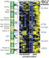
FRQ domains and their time-of-day-specific modifications. A cartoon of the domains of FRQ is shown to the left: CC, coiled-coiled domain; NLS, nuclear localization signal; FCD, FRQ-CKI interacting domain; FFD, FRQ-FRH interacting domain; PEST-#, PEST domains important for protein turnover; frq#, frq alleles followed by period length. Moving to the right in the figure, the positions of phosphorylations are marked. The two heat maps show how these change over time, with yellow marking higher phosphorylation and blue marking lower phosphorylation; each heat map consists of six columns corresponding to 4-h blocks of time in the circadian day from subjective dawn (CT0) through dusk (CT12) to the next night (CT20). The heat map to the right shows total phosphorylation, and, in the heat map to the left, the level of phosphorylation at any time is normalized to the total phosphorylation of the protein to better highlight where changes are occurring; these areas are marked with boxes and arrows showing where they lie within FRQ. Farthest to the right are noted the effects of mutating the serines or threonines that are phosphorylated at the locations noted to alanines. Modified from reference .
FIGURE 5
FIGURE 5
Light resets the clock. (Top) The daily cycle in the abundance of frq RNA is shown along with how a brief exposure to light at any time greatly increases this. (Middle) The cycle of frq transcription is shown in black, and the colored lines show the effect of light-induced frq expression on the steady-state rhythm. (Bottom) The response of the clock to light is juxtaposed with the daily cycle of frq expression: When light is seen while frq levels are rising, the phase of the cycle is advanced because frq rises to peak levels sooner. When light is seen while frq levels are falling, the cycle is set back. Adapted from reference .
FIGURE 6
FIGURE 6
How temperature might reset the circadian cycle. Cycles in FRQ levels at different temperatures are shown in green. A step up in temperature at any time (red arrows) results in not enough FRQ to close the feedback loop, so the clock is reset to the time corresponding to low FRQ, subjective dawn. A step down in temperature at any time leaves the clock with sufficient FRQ to close the feedback loop; the cycle is reset to the time marked by high FRQ, around subjective evening. Adapted from reference .
FIGURE 7
FIGURE 7
Output from the circadian clock. (A) The relationship of output to the oscillator is shown. The circadian clock is fungi and animals can be approximated as a single-step negative feedback loop in which the Positive Elements (in blue, the heterodimer of WC-1 and WC-2 or in animals BMAL1 and CLOCK) drive expression of the gene(s) encoding the Negative Elements (in red). The Negative Elements feed back, physically interact with the Positive Elements, and turn down their activity. The cycle thus generates rhythmic activity of the Positive Elements. When they, in turn, regulate genes whose products do not participate in the feedback loop, there are the means for transcriptional output from the clock. These are the ccgs. Adapted from reference . (B) The abundance of ccgs is shown as a function of the time of day at which their levels peak. The data are from 2-h bins across the day, with a bimodal peak of gene expression in the late night to morning and another peak around dusk (from reference 91).

References

    1. Dunlap JC, Loros JJ, Decoursey P (ed). 2004. Chronobiology: Biological Timekeeping. Sinauer Assoc, Sunderland, MA.
    1. Ingold CT. 1971. Fungal Spores. Clarendon Press, Oxford, United Kingdom.
    1. Bünning E. 1973. The Physiological Clock, 3rd ed. Springer-Verlag, New York, NY. [PubMed]
    1. Brandt WH. 1953. Zonation in a prolineless strain of Neurospora. Mycologia 45:194–208.
    1. Austin B. 1968. An endogenous rhythm of spore discharge in Sordaria fimicola. Ann Bot (Lond) 32:262–278. 10.1093/oxfordjournals.aob.a084207 - DOI

Publication types

MeSH terms

Substances