

Review
doi: 10.1016/j.tins.2017.04.001.
Epub 2017 May 17.
The Role of Astrocyte Dysfunction in Parkinson's Disease Pathogenesis
Affiliations
- PMID: 28527591
- PMCID: PMC5462417
- DOI: 10.1016/j.tins.2017.04.001
Item in Clipboard
Review
The Role of Astrocyte Dysfunction in Parkinson's Disease Pathogenesis
Trends Neurosci.
2017 Jun.
Abstract
Astrocytes are the most populous glial subtype and are critical for brain function. Despite this, historically there have been few studies into the role that they may have in neurodegenerative diseases, such as Parkinson's disease (PD). Recently, however, several studies have determined that genes known to have a causative role in the development of PD are expressed in astrocytes and have important roles in astrocyte function. Here, we review these recent developments and discuss their impact on our understanding of the pathophysiology of PD, and the implications that this might have for its treatment.
Copyright © 2017 The Authors. Published by Elsevier Ltd.. All rights reserved.
Figures
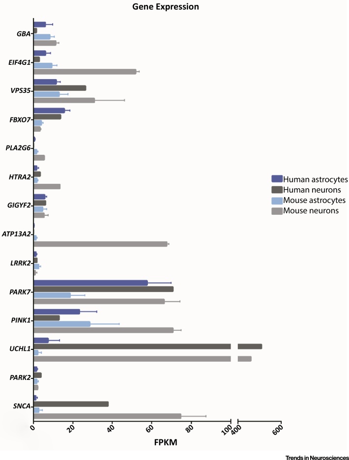
Expression Levels of Key Parkinson’s Disease (PD) Genes in Astrocytes and Neurons. Transcriptome data from Zhang et al. showing the expression levels of genes known to be causative in PD in astrocytes and neurons from humans and mice. Human astrocytes N = 12 subjects; human neurons N = 1 subject; mouse astrocytes N = 6 animals; mouse neurons N = 2 animals. Abbreviation: FPKM, fragments per kilobase of transcript per million mapped reads. Graph shows mean ± SD. Data obtained from Supplementary Table S4 in the original publication , and can be browsed online at http://web.stanford.edu/group/barres_lab/brainseq2/brainseq2.html . For full gene names, please see the main text.
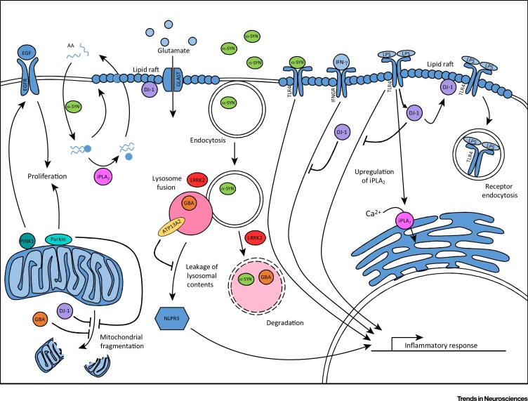
Parkinson’s Disease (PD)-Related Gene Pathways Are Implicated in Astrocytes. Parkin and PINK1 regulate proliferation, which, in the case of PINK1, occurs through the EGF signalling pathway. PINK1, Parkin, GBA, and DJ-1 have all been shown to have a role in the maintenance of healthy mitochondria. Astrocyte α-synuclein (α-SYN) regulates the uptake and distribution of arachidonic acid (AA), which is released from phospholipids by Group VI Ca2+-independent phospholipase A2 (iPLA2). DJ-1 regulates the stability of lipid rafts, therefore maintaining the glutamate transporter GLAST at the membrane. Additionally, DJ-1 is involved in the termination of TLR4 signalling via receptor endocytosis, and the inhibition of the IFN-γ inflammatory response. iPLA2 is upregulated in response to TLR4 signalling and increases the calcium load in the endoplasmic reticulum. Extracellular α-SYN can be endocytosed via a TLR4-independent process and degraded by the lysosome. At high concentrations, extracellular α-SYN activates TLR4 signalling that is not terminated by receptor endocytosis. GBA mutations can disrupt degradation of proteins via the autophagy pathway. Leucine-rich repeat kinase 2 (LRRK2) regulates fusion and/or degradation in the autophagy pathway and, alongside lysosomal type 5 ATPase (ATP13A2), may have a role in the control of lysosomal pH. ATP13A2 levels maintain the stability of the lysosome and prevent its contents from leaking into the cytosol, which can result in activation of the NLPR3 inflammasome. Abbreviation: LPS, lipopolysaccharide.
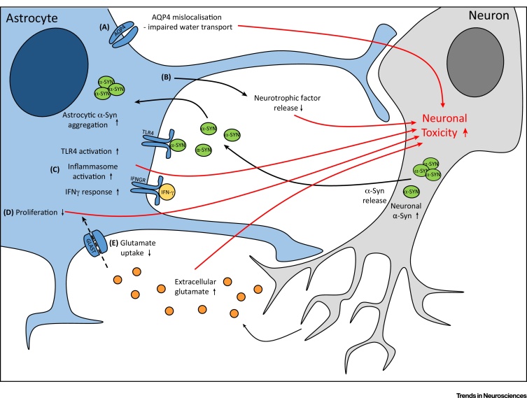
Dysfunctional Astrocytes Contribute to Neuronal Toxicity. Astrocyte dysfunction elicits neuronal toxicity via five main mechanisms. (A) Aquaporin-4 (AQP4) water channels are mislocalised away from the astrocyte end-feet, resulting in impaired water transport. (B) The neuroprotective capacity of astrocytes is reduced because of decreased neurotrophic factor release. (C) Inflammatory signalling via the TLR4, IFN-γ, and NLPR3 inflammasome pathways is increased. (D) Astrocyte proliferation is impaired, reducing the capacity of the cells to respond to an insult. (E) Glutamate uptake is reduced, potentially resulting in increased extracellular glutamate and, therefore, neuronal excitotoxicity. Abbreviation: α-SYN, α- synuclein.
References
-
- Miklossy J. Role of ICAM-1 in persisting inflammation in Parkinson disease and MPTP monkeys. Exp. Neurol. 2006;197:275–283. - PubMed
Publication types
MeSH terms
Grants and funding
LinkOut - more resources
Full Text Sources
Other Literature Sources
Medical
