

A machine-learning graph-based approach for 3D segmentation of Bruch's membrane opening from glaucomatous SD-OCT volumes
- PMID: 28528295
- PMCID: PMC5729043
- DOI: 10.1016/j.media.2017.04.007
A machine-learning graph-based approach for 3D segmentation of Bruch's membrane opening from glaucomatous SD-OCT volumes
Abstract
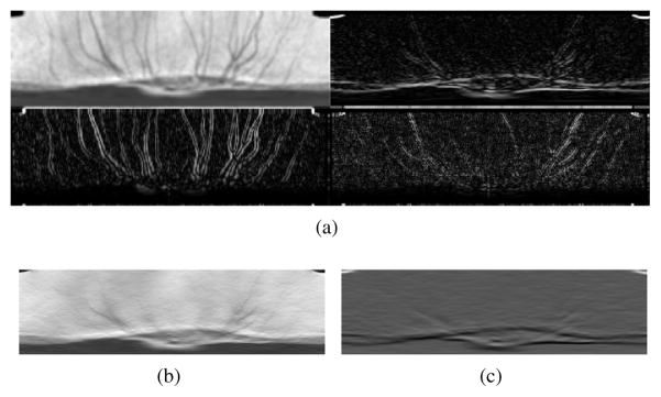
Bruch's membrane opening-minimum rim width (BMO-MRW) is a recently proposed structural parameter which estimates the remaining nerve fiber bundles in the retina and is superior to other conventional structural parameters for diagnosing glaucoma. Measuring this structural parameter requires identification of BMO locations within spectral domain-optical coherence tomography (SD-OCT) volumes. While most automated approaches for segmentation of the BMO either segment the 2D projection of BMO points or identify BMO points in individual B-scans, in this work, we propose a machine-learning graph-based approach for true 3D segmentation of BMO from glaucomatous SD-OCT volumes. The problem is formulated as an optimization problem for finding a 3D path within the SD-OCT volume. In particular, the SD-OCT volumes are transferred to the radial domain where the closed loop BMO points in the original volume form a path within the radial volume. The estimated location of BMO points in 3D are identified by finding the projected location of BMO points using a graph-theoretic approach and mapping the projected locations onto the Bruch's membrane (BM) surface. Dynamic programming is employed in order to find the 3D BMO locations as the minimum-cost path within the volume. In order to compute the cost function needed for finding the minimum-cost path, a random forest classifier is utilized to learn a BMO model, obtained by extracting intensity features from the volumes in the training set, and computing the required 3D cost function. The proposed method is tested on 44 glaucoma patients and evaluated using manual delineations. Results show that the proposed method successfully identifies the 3D BMO locations and has significantly smaller errors compared to the existing 3D BMO identification approaches.
Keywords: Bruch’s membrane opening; Ophthalmology; Optic disc; Retina; SD-OCT; Segmentation.
Published by Elsevier B.V.
Figures


Similar articles
-
Novel Bruch's Membrane Opening Minimum Rim Area Equalizes Disc Size Dependency and Offers High Diagnostic Power for Glaucoma.Invest Ophthalmol Vis Sci. 2016 Dec 1;57(15):6596-6603. doi: 10.1167/iovs.16-20561. Invest Ophthalmol Vis Sci. 2016. PMID: 27951592
-
Comparing optical coherence tomography radial and cube scan patterns for measuring Bruch's membrane opening minimum rim width (BMO-MRW) in glaucoma and healthy eyes: cross-sectional and longitudinal analysis.Br J Ophthalmol. 2018 Mar;102(3):344-351. doi: 10.1136/bjophthalmol-2016-310111. Epub 2017 Aug 3. Br J Ophthalmol. 2018. PMID: 28774935
-
Incorporation of gradient vector flow field in a multimodal graph-theoretic approach for segmenting the internal limiting membrane from glaucomatous optic nerve head-centered SD-OCT volumes.Comput Med Imaging Graph. 2017 Jan;55:87-94. doi: 10.1016/j.compmedimag.2016.06.007. Epub 2016 Jul 25. Comput Med Imaging Graph. 2017. PMID: 27507325 Free PMC article.
-
Glaucoma Diagnosis of the Optic Nerve Head Using Optical Coherence Tomography - Significance of Bruch's Membrane Opening and Derived OCT Parameters.Klin Monbl Augenheilkd. 2025 Jul;242(7):718-725. doi: 10.1055/a-2558-8721. Epub 2025 Jul 24. Klin Monbl Augenheilkd. 2025. PMID: 40719088 Review. English, German.
-
[Correlation between Lamina Cribrosa Tilt, Myopia and Glaucoma Using Optical Coherence Tomography with a Wide Band Femtosecond Mode-locked Laser].Nippon Ganka Gakkai Zasshi. 2016 Nov;120(11):764-71. Nippon Ganka Gakkai Zasshi. 2016. PMID: 30074741 Review. Japanese.
Cited by
-
Automated detection of exudative age-related macular degeneration in spectral domain optical coherence tomography using deep learning.Graefes Arch Clin Exp Ophthalmol. 2018 Feb;256(2):259-265. doi: 10.1007/s00417-017-3850-3. Epub 2017 Nov 20. Graefes Arch Clin Exp Ophthalmol. 2018. PMID: 29159541
-
The Future of Imaging in Detecting Glaucoma Progression.Ophthalmology. 2017 Dec;124(12S):S76-S82. doi: 10.1016/j.ophtha.2017.10.011. Ophthalmology. 2017. PMID: 29157365 Free PMC article. Review.
-
DRUNET: a dilated-residual U-Net deep learning network to segment optic nerve head tissues in optical coherence tomography images.Biomed Opt Express. 2018 Jun 25;9(7):3244-3265. doi: 10.1364/BOE.9.003244. eCollection 2018 Jul 1. Biomed Opt Express. 2018. PMID: 29984096 Free PMC article.
-
An Automated CAD System for Accurate Grading of Uveitis Using Optical Coherence Tomography Images.Sensors (Basel). 2021 Aug 13;21(16):5457. doi: 10.3390/s21165457. Sensors (Basel). 2021. PMID: 34450898 Free PMC article.
-
Weakly supervised individual ganglion cell segmentation from adaptive optics OCT images for glaucomatous damage assessment.Optica. 2021 May 20;8(5):642-651. doi: 10.1364/optica.418274. Epub 2021 May 4. Optica. 2021. PMID: 35174258 Free PMC article.
References
MeSH terms
Grants and funding
LinkOut - more resources
Full Text Sources
Other Literature Sources
Medical
