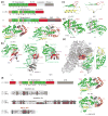
The group II intron maturase: a reverse transcriptase and splicing factor go hand in hand
- PMID: 28528306
- PMCID: PMC5694389
- DOI: 10.1016/j.sbi.2017.05.002
The group II intron maturase: a reverse transcriptase and splicing factor go hand in hand
Abstract
The splicing of group II introns in vivo requires the assistance of a multifunctional intron encoded protein (IEP, or maturase). Each IEP is also a reverse-transcriptase enzyme that enables group II introns to behave as mobile genetic elements. During splicing or retro-transposition, each group II intron forms a tight, specific complex with its own encoded IEP, resulting in a highly reactive holoenzyme. This review focuses on the structural basis for IEP function, as revealed by recent crystal structures of an IEP reverse transcriptase domain and cryo-EM structures of an IEP-intron complex. These structures explain how the same IEP scaffold is utilized for intron recognition, splicing and reverse transcription, while providing a physical basis for understanding the evolutionary transformation of the IEP into the eukaryotic splicing factor Prp8.
Copyright © 2017 Elsevier Ltd. All rights reserved.
Conflict of interest statement
The authors declare no competing financial interests.
Figures


Similar articles
-
A group II intron-encoded maturase functions preferentially in cis and requires both the reverse transcriptase and X domains to promote RNA splicing.J Mol Biol. 2004 Jul 2;340(2):211-31. doi: 10.1016/j.jmb.2004.05.004. J Mol Biol. 2004. PMID: 15201048
-
The brown algae Pl.LSU/2 group II intron-encoded protein has functional reverse transcriptase and maturase activities.PLoS One. 2013;8(3):e58263. doi: 10.1371/journal.pone.0058263. Epub 2013 Mar 11. PLoS One. 2013. PMID: 23505475 Free PMC article.
-
Structure of a Thermostable Group II Intron Reverse Transcriptase with Template-Primer and Its Functional and Evolutionary Implications.Mol Cell. 2017 Dec 7;68(5):926-939.e4. doi: 10.1016/j.molcel.2017.10.024. Epub 2017 Nov 16. Mol Cell. 2017. PMID: 29153391 Free PMC article.
-
Insights into the strategies used by related group II introns to adapt successfully for the colonisation of a bacterial genome.RNA Biol. 2014;11(8):1061-71. doi: 10.4161/rna.32092. Epub 2014 Oct 31. RNA Biol. 2014. PMID: 25482895 Free PMC article. Review.
-
Group II introns: structure and catalytic versatility of large natural ribozymes.Crit Rev Biochem Mol Biol. 2003;38(3):249-303. doi: 10.1080/713609236. Crit Rev Biochem Mol Biol. 2003. PMID: 12870716 Review.
Cited by
-
Exon and protein positioning in a pre-catalytic group II intron RNP primed for splicing.Nucleic Acids Res. 2020 Nov 4;48(19):11185-11198. doi: 10.1093/nar/gkaa773. Nucleic Acids Res. 2020. PMID: 33021674 Free PMC article.
-
U5 snRNA Interactions With Exons Ensure Splicing Precision.Front Genet. 2021 Jul 2;12:676971. doi: 10.3389/fgene.2021.676971. eCollection 2021. Front Genet. 2021. PMID: 34276781 Free PMC article.
-
Research Progress of Group II Intron Splicing Factors in Land Plant Mitochondria.Genes (Basel). 2024 Jan 28;15(2):176. doi: 10.3390/genes15020176. Genes (Basel). 2024. PMID: 38397166 Free PMC article. Review.
-
An Allosteric Network for Spliceosome Activation Revealed by High-Throughput Suppressor Analysis in Saccharomyces cerevisiae.Genetics. 2019 May;212(1):111-124. doi: 10.1534/genetics.119.301922. Epub 2019 Mar 21. Genetics. 2019. PMID: 30898770 Free PMC article.
-
Organellar Introns in Fungi, Algae, and Plants.Cells. 2021 Aug 6;10(8):2001. doi: 10.3390/cells10082001. Cells. 2021. PMID: 34440770 Free PMC article. Review.
References
-
- Peebles CL, Perlman PS, Mecklenburg KL, Petrillo ML, Tabor JH, Jarrell KA, Cheng HL. A self-splicing RNA excises an intron lariat. Cell. 1986;44:213–223. - PubMed
Publication types
MeSH terms
Substances
Grants and funding
LinkOut - more resources
Full Text Sources
Other Literature Sources

