

MicroRNA-30a-5p (miR-30a) regulates cell motility and EMT by directly targeting oncogenic TM4SF1 in colorectal cancer
- PMID: 28528497
- PMCID: PMC11819409
- DOI: 10.1007/s00432-017-2440-4
MicroRNA-30a-5p (miR-30a) regulates cell motility and EMT by directly targeting oncogenic TM4SF1 in colorectal cancer
Abstract
Purpose: Colorectal cancer (CRC) is one of the leading causes of cancer death worldwide, and many oncogenes and tumor suppressor genes are involved in CRC. MicroRNAs (miRNAs) are small non-coding RNAs that can negatively regulate gene expression. Previous studies have revealed that miRNAs regulate the development and progression of many cancers. In this study, we investigated the role of microRNA-30a-5p (miR-30a) in CRC and its unknown mechanisms.
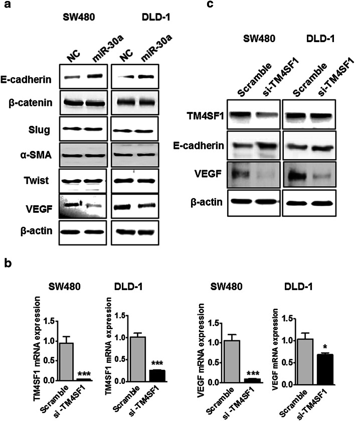
Methods: qRT-PCR was used to detect miR-30a and TM4SF1 mRNA expression in CRC specimens and cell lines. CRC cell migration and invasion were assessed after transfection with miR-30a or TM4SF1 using wound healing and trans-well migration and invasion assays. Transmembrane-4-L-six-family protein (TM4SF1) was validated as a target of miR-30a in CRC through luciferase reporter assay and bioinformatics algorithms. Moreover, two EMT regulators, E-cadherin and VEGF, were also identified using Western blotting and immunohistochemistry.
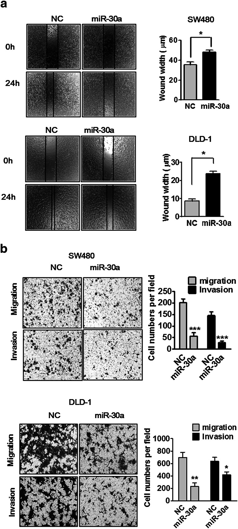
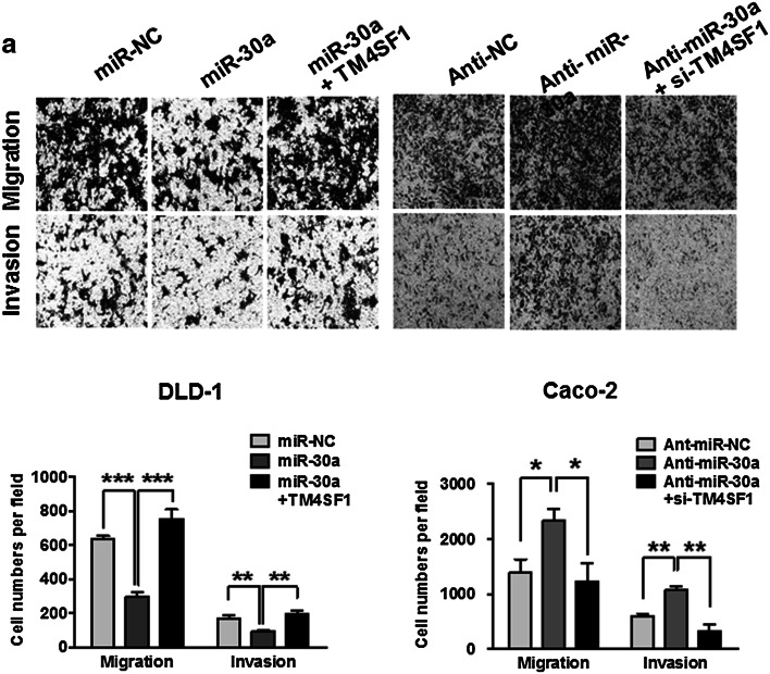
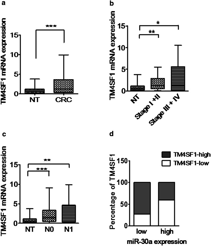
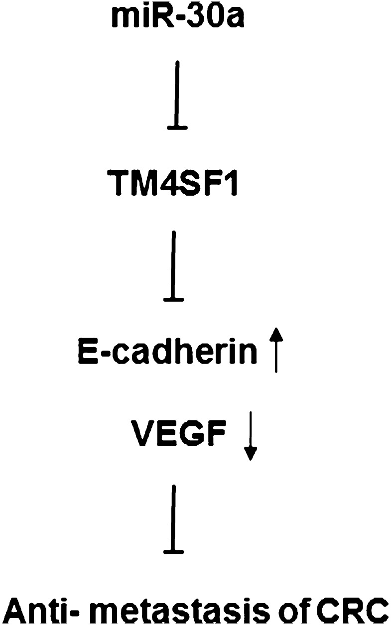
Results: We found that miR-30a was down-regulated in CRC tumor tissues and cell lines, and miR-30a was inversely associated with advanced stage and lymph node metastatic status compared with normal tissues. miR-30a decreased migration and invasion in CRC cell lines, and miR-30a overexpression not only down-regulated TM4SF1 mRNA and protein expression, but also inhibited the expression of VEGF and enhanced expression of E-cadherin. We also showed that TM4SF1 was up-regulated in CRC tumor specimens compared with adjacent normal tissues, and TM4SF1 expression was significantly associated with advanced stage and lymph node status compared with adjacent normal tissues.
Conclusions: These results suggest that miR-30a is an important regulator of TM4SF1, VEGF, and E-cadherin for CRC lymph node metastasis, a potential new therapeutic target in CRC.
Keywords: Colorectal cancer; Metastasis; Transmembrane-4-L-six-family protein 1; miR-30a.
Conflict of interest statement
The authors declare that they have no competing interests.
Figures


Similar articles
-
MicroRNA-9 suppresses cell migration and invasion through downregulation of TM4SF1 in colorectal cancer.Int J Oncol. 2016 May;48(5):2135-43. doi: 10.3892/ijo.2016.3430. Epub 2016 Mar 9. Int J Oncol. 2016. PMID: 26983891
-
Colorectal cancer-secreted exosomal circ_001422 plays a role in regulating KDR expression and activating mTOR signaling in endothelial cells by targeting miR-195-5p.J Cancer Res Clin Oncol. 2023 Oct;149(13):12227-12240. doi: 10.1007/s00432-023-05095-1. Epub 2023 Jul 11. J Cancer Res Clin Oncol. 2023. PMID: 37432457 Free PMC article.
-
MiRNA-206 suppresses PGE2-induced colorectal cancer cell proliferation, migration, and invasion by targetting TM4SF1.Biosci Rep. 2018 Sep 19;38(5):BSR20180664. doi: 10.1042/BSR20180664. Print 2018 Oct 31. Biosci Rep. 2018. PMID: 30135139 Free PMC article.
-
MicroRNAs that regulate PTEN as potential biomarkers in colorectal cancer: a systematic review.J Cancer Res Clin Oncol. 2020 Apr;146(4):809-820. doi: 10.1007/s00432-020-03172-3. Epub 2020 Mar 7. J Cancer Res Clin Oncol. 2020. PMID: 32146564 Free PMC article.
-
Role of Mesenchymal Markers in Colorectal Cancer Metastasis.Mol Biol Rep. 2025 Jul 4;52(1):673. doi: 10.1007/s11033-025-10745-3. Mol Biol Rep. 2025. PMID: 40613936 Review.
Cited by
-
Tumor cell stemness in gastrointestinal cancer: regulation and targeted therapy.Front Mol Biosci. 2024 Feb 22;10:1297611. doi: 10.3389/fmolb.2023.1297611. eCollection 2023. Front Mol Biosci. 2024. PMID: 38455361 Free PMC article. Review.
-
LDR-Induced miR-30a and miR-30b Target the PAI-1 Pathway to Control Adverse Effects of NSCLC Radiotherapy.Mol Ther. 2019 Feb 6;27(2):342-354. doi: 10.1016/j.ymthe.2018.10.015. Epub 2018 Oct 26. Mol Ther. 2019. PMID: 30424954 Free PMC article.
-
Colorectal liver metastasis: molecular mechanism and interventional therapy.Signal Transduct Target Ther. 2022 Mar 4;7(1):70. doi: 10.1038/s41392-022-00922-2. Signal Transduct Target Ther. 2022. PMID: 35246503 Free PMC article. Review.
-
Long noncoding RNA NORAD regulates lung cancer cell proliferation, apoptosis, migration, and invasion by the miR-30a-5p/ADAM19 axis.Int J Clin Exp Pathol. 2020 Jan 1;13(1):1-13. eCollection 2020. Int J Clin Exp Pathol. 2020. PMID: 32055266 Free PMC article.
-
TM4SF1 promotes EMT and cancer stemness via the Wnt/β-catenin/SOX2 pathway in colorectal cancer.J Exp Clin Cancer Res. 2020 Nov 5;39(1):232. doi: 10.1186/s13046-020-01690-z. J Exp Clin Cancer Res. 2020. PMID: 33153498 Free PMC article.
References
-
- Allioli N, Vincent S, Vlaeminck-Guillem V, Decaussin-Petrucci M, Ragage F, Ruffion A, Samarut J (2011) TM4SF1, a novel primary androgen receptor target gene over-expressed in human prostate cancer and involved in cell migration. Prostate 71(11):1239–1250 - PubMed
-
- Baraniskin A, Birkenkamp-Demtroder K, Maghnouj A, Zollner H, Munding J, Klein-Scory S, Reinacher-Schick A, Schwarte-Waldhoff I, Schmiegel W, Hahn SA (2012) MiR-30a-5p suppresses tumor growth in colon carcinoma by targeting DTL. Carcinogenesis 33(4):732–739 - PubMed
-
- Bartel DP (2004) MicroRNAs: genomics, biogenesis, mechanism, and function. Cell 116(2):281–297 - PubMed
-
- Brenner H, Kloor M, Pox CP (2014) Colorectal cancer. Lancet 383(9927):1490–1502 - PubMed
MeSH terms
Substances
LinkOut - more resources
Full Text Sources
Other Literature Sources
Medical
Research Materials
Miscellaneous
