

Review
doi: 10.1016/j.mrrev.2016.07.002.
Epub 2016 Jul 5.
Human papillomavirus molecular biology
Affiliations
- PMID: 28528688
- PMCID: PMC5500221
- DOI: 10.1016/j.mrrev.2016.07.002
Item in Clipboard
Review
Human papillomavirus molecular biology
Mutat Res Rev Mutat Res.
2017 Apr-Jun.
Abstract
Human papillomaviruses are small DNA viruses with a tropism for squamous epithelia. A unique aspect of human papillomavirus molecular biology involves dependence on the differentiation status of the host epithelial cell to complete the viral lifecycle. A small group of these viruses are the etiologic agents of several types of human cancers, including oral and anogenital tract carcinomas. This review focuses on the basic molecular biology of human papillomaviruses.
Keywords: Cervical cancer; Epithelial differentiation; Human papillomavirus; Oncogene; Tumor suppressor; Vaccine.
Copyright © 2016 Elsevier B.V. All rights reserved.
Figures
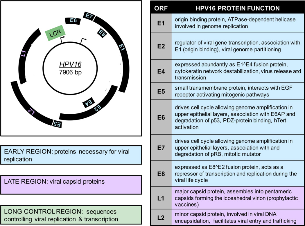
On the left, the HPV16 genomic map of 7906 base pairs is shown. Only the coding strand is included and transcription occurs in the clockwise direction. The early promoter (P97) is indicated by an arrow at the approximate position of the RNA initiation site in the long control region LCR. The late promoter (P670) is also indicated by an arrow at its initiation site in the E7 ORF. The early region is depicted in blue and contains proteins necessary for viral replication including E1, E2, E3, E4, E5, E6 and E7. The late region is shown in purple and contains the viral capsid proteins L1 and L2. The LCR is shown in green and contains sequences controlling viral replication & transcription. On the right, a table of the HPV16 ORFs and a brief description of their corresponding viral functions is shown. More details can be found in section 1.3 of the text.
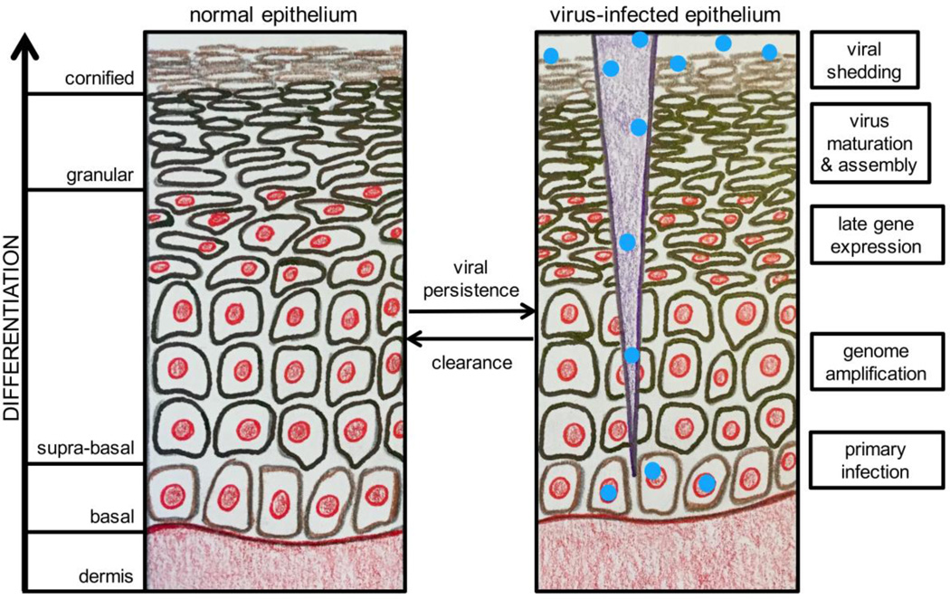
An illustration of normal differentiating squamous epithelium is shown on the left with the layers of the differentiating epithelium noted. On the right, a brief description of the HPV life cycle stage occurring in the corresponding epithelial layer is shown. Greater detail on HPV productive infection and the viral life cycle is included in section 1.5. This figure was illustrated by M. E. Harden and adapted from a figure by C. L. Nguyen.
References
-
- CDC. Epidemiology and Prevention of Vaccine-Preventable Diseases. 13. Washington, D.C.: Public Health Foundation; 2015.
-
- Schiffman M, Castle PE, Jeronimo J, Rodriguez AC, Wacholder S. Human papillomavirus and cervical cancer. Lancet. 2007;370:890–907. - PubMed
-
- Bray F, Ren JS, Masuyer E, Ferlay J. Global estimates of cancer prevalence for 27 sites in the adult population in 2008. Int J Cancer. 2013;132:1133–1145. - PubMed
-
- Forman D, de Martel C, Lacey CJ, Soerjomataram I, Lortet-Tieulent J, Bruni L, Vignat J, Ferlay J, Bray F, Plummer M, Franceschi S. Global burden of human papillomavirus and related diseases. Vaccine. 2012;(30 Suppl 5):F12–F23. - PubMed
Publication types
MeSH terms
Substances
Grants and funding
LinkOut - more resources
Full Text Sources
Other Literature Sources
