An official website of the United States government
The .gov means it’s official.
Federal government websites often end in .gov or .mil. Before
sharing sensitive information, make sure you’re on a federal
government site.
The site is secure.
The https:// ensures that you are connecting to the
official website and that any information you provide is encrypted
and transmitted securely.
1 Department of Molecular, Cell, and Cancer Biology, University of Massachusetts Medical School, Worcester, MA 01605, USA.
2 Department of Molecular, Cellular, and Biomedical Sciences, University of New Hampshire, Durham, NH 03824, USA.
3 Department of Molecular, Cell, and Cancer Biology, University of Massachusetts Medical School, Worcester, MA 01605, USA; Howard Hughes Medical Institute, University of Massachusetts Medical School, Worcester, MA 01605, USA.
4 Department of Molecular, Cell, and Cancer Biology, University of Massachusetts Medical School, Worcester, MA 01605, USA. Electronic address: thomas.fazzio@umassmed.edu.
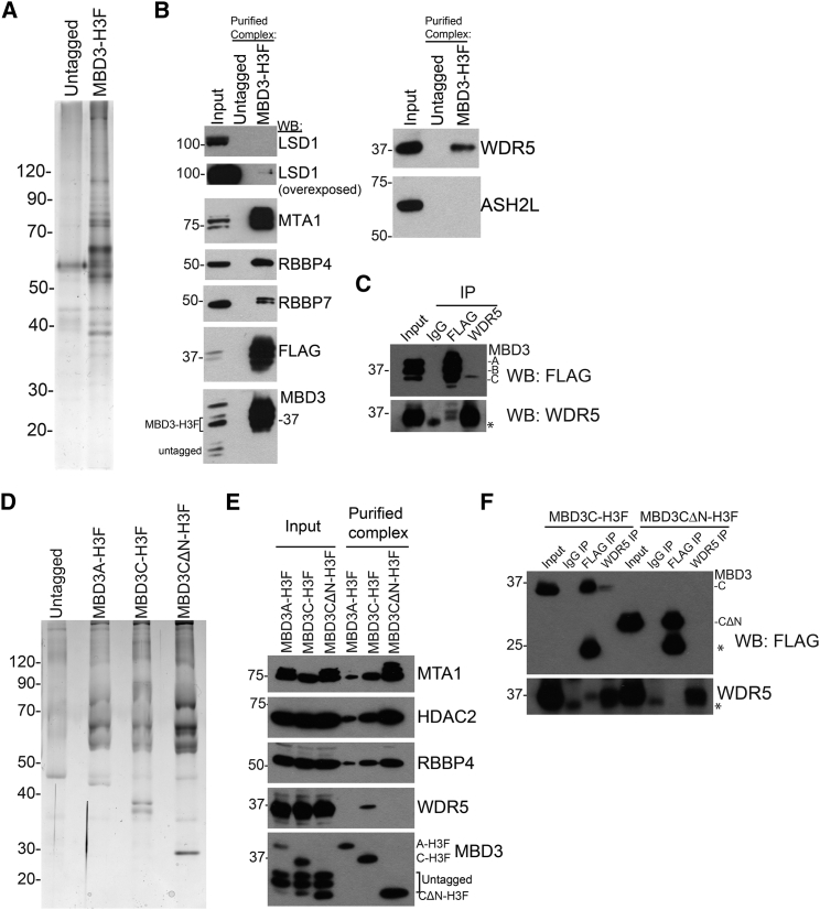
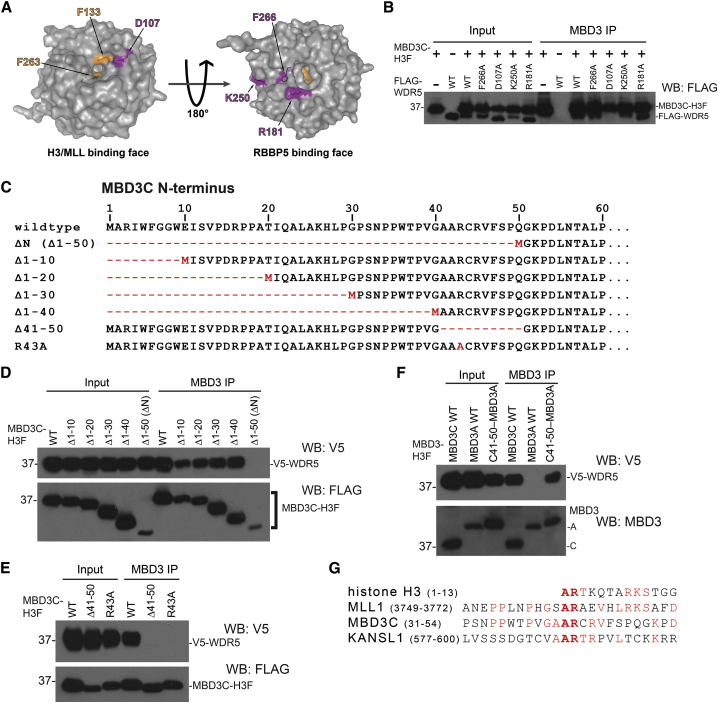
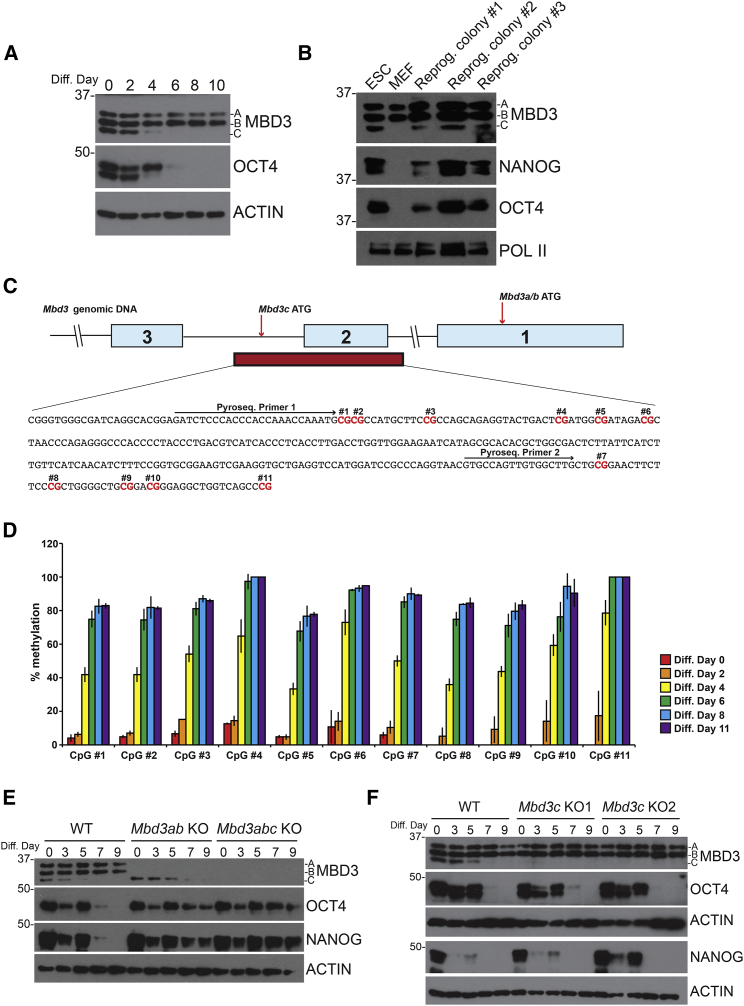
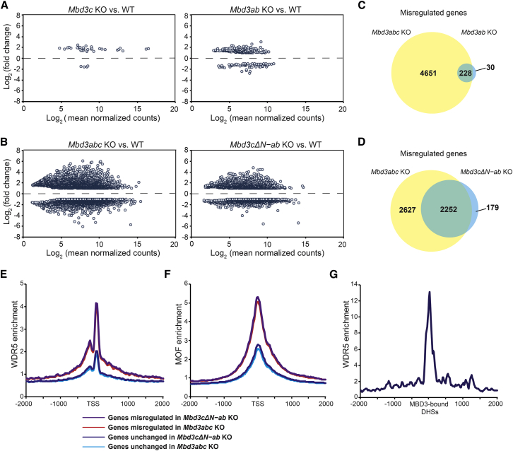
The Nucleosome Remodeling and Deacetylase (NuRD) complex is a chromatin regulatory complex that functions as a transcriptional co-repressor in metazoans. The NuRD subunit MBD3 is essential for targeting and assembly of a functional NuRD complex as well as embryonic stem cell (ESC) pluripotency. Three MBD3 isoforms (MBD3A, MBD3B, and MBD3C) are expressed in mouse. Here, we find that the MBD3C isoform contains a unique 50-amino-acid N-terminal region that is necessary for MBD3C to specifically interact with the histone H3 binding protein WDR5. Domain analyses of WDR5 reveal that the H3 binding pocket is required for interaction with MBD3C. We find that while Mbd3c knockout ESCs differentiate normally, MBD3C is redundant with the MBD3A and MBD3B isoforms in regulation of gene expression, with the unique MBD3C N terminus required for this redundancy. Together, our data characterize a unique NuRD complex variant that functions specifically in ESCs.
The NuRD Subunit MBD3C Co-purifies with WDR5 (A) Silver stain of MBD3-H3F complex.…
Figure 1
The NuRD Subunit MBD3C Co-purifies with WDR5 (A) Silver stain of MBD3-H3F complex. (B) Western blot (WB) of purified complex from (A) showing interaction of MBD3 with NuRD subunits (left) and with WDR5 or MLL subunit ASH2L (right). (C) Western blots for MBD3-H3F or WDR5 upon immunoprecipitation (IP) of each. Asterisk indicates immunoglobulin G (IgG). (D) Silver stain of MBD3 complex expressing individually H3F-tagged MBD3 isoforms. (E) Western blot of purified complexes from (D) showing interaction with WDR5 and NuRD subunits. (F) WDR5 or FLAG immunoprecipitation from individually tagged MBD3C-H3F and MBD3CΔN-H3F ESCs. Asterisks indicate IgG.
Figure 2
The WDR5 Histone H3 Binding…
Figure 2
The WDR5 Histone H3 Binding Pocket Is Required to Bind MBD3C (A) PyMol…
Figure 2
The WDR5 Histone H3 Binding Pocket Is Required to Bind MBD3C (A) PyMol depiction of WDR5 crystal structure (PDB: 2GNQ) showing the H3K4/MLL1 (left) and RBBP5 (right) binding pockets. Residues individually mutated to alanine (Yang et al., 2014) are shown in magenta. Residues necessary for MLL1 R3761 or histone H3 R2 binding are shown in orange. (B) CoIPs with MBD3 antibody from 293T cells co-transfected with expression vectors carrying MBD3C-H3F and indicated FLAG-tagged WDR5 mutants. (C) Schematic of Mbd3c N-terminal mutant constructs used in (D) and (E). (D–F) CoIPs with MBD3 antibody in 293T cells performed as in (B), using V5-tagged WDR5 constructs and H3F-tagged MBD3 constructs. For “C41-50−MBD3A,” MBD3C amino acids 41–50 were fused to the N terminus of the MBD3A isoform. (G) Alignment of the MBD3C N terminus with WDR5-binding regions of mouse MLL1, KANSL1, and histone H3.
Figure 3
Mbd3c Is Dispensable for ESC…
Figure 3
Mbd3c Is Dispensable for ESC Differentiation (A) Western blots from ESCs differentiated over…
Figure 3
Mbd3c Is Dispensable for ESC Differentiation (A) Western blots from ESCs differentiated over 10 days. Actin serves as a loading control. (B) Western blots from primary MEFs reprogrammed to iPSCs. (C) Schematic of the Mbd3 gene showing the sequence and location of the Mbd3c promoter CpG island (red bar). Light-blue boxes indicate exons. CpGs tested for methylation are highlighted in red. (D) Pyrosequencing of bisulfite-converted DNA from cells collected at the indicated differentiation time points. Error bars represent the SD of three biological replicates. (E and F) Western blots of differentiating Mbd3ab and Mbd3abc KO cells (E) and two clonal Mbd3c KO lines (F). WT, wild-type.
Figure 4
MBD3C Is Redundant with MBD3A…
Figure 4
MBD3C Is Redundant with MBD3A and MBD3B in Regulation of Gene Expression (A…
Figure 4
MBD3C Is Redundant with MBD3A and MBD3B in Regulation of Gene Expression (A and B) MA plots showing log2 (fold change) in gene expression in Mbd3c KO (A, left), Mbd3ab KO (A, right), Mbd3abc KO (B, left), and Mbd3cΔN-ab KO (B, right) ESCs relative to wild-type (WT). Genes shown are misregulated ≥2-fold compared with WT. (C and D) Venn diagrams showing overlap between misregulated genes in ESCs of indicated genotypes. (E and F) WDR5 binding (Ang et al., 2011) (E) and MOF binding (Li et al., 2012) (F) averaged over transcription start sites (TSS) of misregulated or unchanged genes in Mbd3abc KO (red) and Mbd3cΔN-ab KO ESCs (purple). (G) Average WDR5 binding over MBD3-bound (Yildirim et al., 2011), TSS-distal DNase I hypersensitive sites (DHSs) (GSM1014514).
Ang Y.S., Tsai S.Y., Lee D.F., Monk J., Su J., Ratnakumar K., Ding J., Ge Y., Darr H., Chang B. Wdr5 mediates self-renewal and reprogramming via the embryonic stem cell core transcriptional network. Cell. 2011;145:183–197.
-
PMC
-
PubMed
Bernstein B.E., Humphrey E.L., Erlich R.L., Schneider R., Bouman P., Liu J.S., Kouzarides T., Schreiber S.L. Methylation of histone H3 Lys 4 in coding regions of active genes. Proc. Natl. Acad. Sci. USA. 2002;99:8695–8700.
-
PMC
-
PubMed
Bode D., Yu L., Tate P., Pardo M., Choudhary J. Characterization of two distinct nucleosome remodeling and deacetylase (NuRD) complex assemblies in embryonic stem cells. Mol. Cell. Proteomics. 2016;15:878–891.
-
PMC
-
PubMed
Chen P.B., Hung J.H., Hickman T.L., Coles A.H., Carey J.F., Weng Z., Chu F., Fazzio T.G. Hdac6 regulates Tip60-p400 function in stem cells. Elife. 2013;2:e01557.
-
PMC
-
PubMed
Cong L., Ran F.A., Cox D., Lin S., Barretto R., Habib N., Hsu P.D., Wu X., Jiang W., Marraffini L.A. Multiplex genome engineering using CRISPR/Cas systems. Science. 2013;339:819–823.
-
PMC
-
PubMed