. 2017 Jun 6;8(6):1516-1524.
doi: 10.1016/j.stemcr.2017.04.023. Epub 2017 May 18.

Directed Differentiation of Human Pluripotent Stem Cells to Microglia

Affiliations

Directed Differentiation of Human Pluripotent Stem Cells to Microglia

Panagiotis Douvaras et al. Stem Cell Reports. .

Abstract

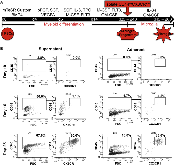
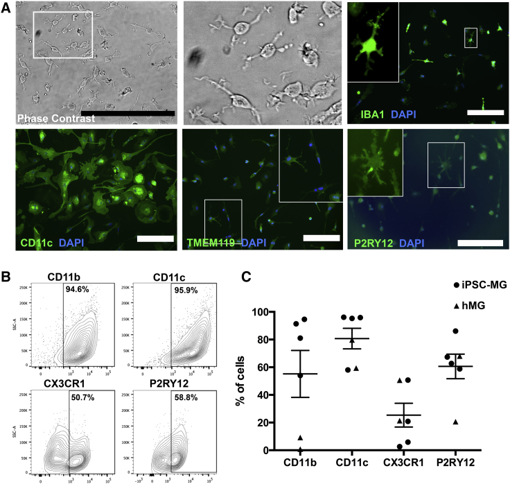
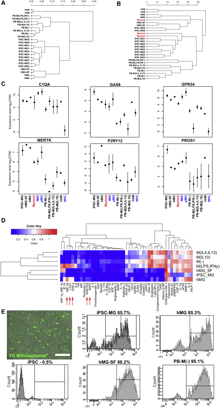
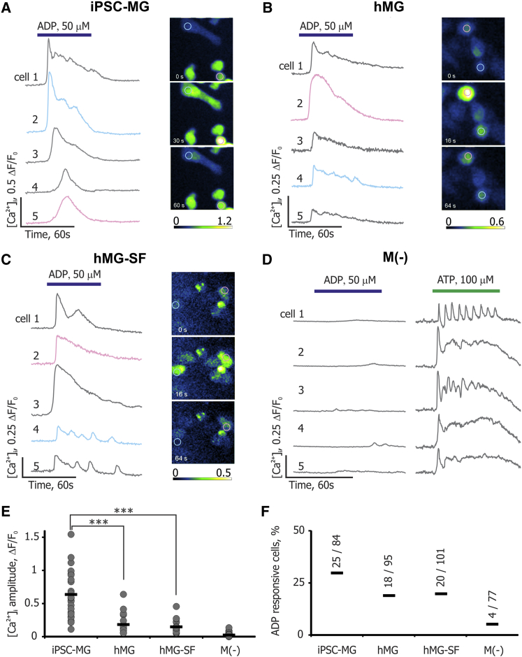
Microglia, the immune cells of the brain, are crucial to proper development and maintenance of the CNS, and their involvement in numerous neurological disorders is increasingly being recognized. To improve our understanding of human microglial biology, we devised a chemically defined protocol to generate human microglia from pluripotent stem cells. Myeloid progenitors expressing CD14/CX3CR1 were generated within 30 days of differentiation from both embryonic and induced pluripotent stem cells (iPSCs). Further differentiation of the progenitors resulted in ramified microglia with highly motile processes, expressing typical microglial markers. Analyses of gene expression and cytokine release showed close similarities between iPSC-derived (iPSC-MG) and human primary microglia as well as clear distinctions from macrophages. iPSC-MG were able to phagocytose and responded to ADP by producing intracellular Ca2+ transients, whereas macrophages lacked such response. The differentiation protocol was highly reproducible across several pluripotent stem cell lines.

Keywords: human microglia; human pluripotent stem cells; microglial differentiation.


Figures

Figure 1
Figure 1
PSC Differentiate to Microglia through Myeloid Progenitors (A) Diagram depicting the major steps of the microglial differentiation protocol. (B) Kinetics of CD45, CX3CR1, and CD14 expression between day 10 and day 25 in the adherent and supernatant fractions. See also Figure S1.
Figure 2
Figure 2
Characterization of iPSC-MG (A) Panel of representative images of iPSC-MG in phase contrast and after immunofluorescent labeling for IBA1, CD11c, TMEM119, and P2RY12. White boxes indicate the areas of the magnified insets. Scale bars, 50 μm (phase contrast) and 200 μm (all other images). (B) Representative flow-cytometry plots for typical microglial surface antigens in iPSC-MG. (C) Dot plot showing the percentage of total cells expressing the microglial surface antigens shown in (B) across four independent iPSC-MG (depicted as circles) and two hMG samples (depicted as triangles). Error bars denote mean ± SEM. See also Movie S1 and Figure S2 for characterization of hMG.
Figure 3
Figure 3
Gene Expression, Cytokine Release Profile, and Phagocytosis of Microglia and Macrophages (A) Hierarchical clustering dendrogram of the RNA-seq data based on global mRNA expression. Sample distances were calculated from Pearson's correlation coefficient. (B) Dendrogram showing hierarchical clustering of our RNA-seq data and data obtained from an independent study of human primary CD45+ cells in the brain (“Myeloid” samples in red, GEO: GSE73721). Analysis is based on transcriptome-wide expression. (C) Graphs showing the expression levels of the six human microglial signature genes. Error bars are means ± SD. Colored samples correspond to data from independent studies (GEO: GSE73721 in red; GEO: GSE85839 in blue). (D) Heatmap of the released cytokine profiles of five independent iPSC-MG runs from two lines, two independent hMG samples, and one hMG-SF sample compared with PB-M. Red arrows indicate the five proteins upregulated in hMG and M(LPS,IFNγ). (E) Representative fluorescent image and flow-cytometry histograms showing phagocytosis of yellow-green (YG)-labeled microspheres. Scale bar, 200 μm. iPSC, undifferentiated iPSCs used as negative control. See also Figure S3; Tables S1 and S2.
Figure 4
Figure 4
ADP-Evoked [Ca2+]i Transients in Microglia and Macrophages (A) Left panel shows five example traces of [Ca2+]i transients following ADP application in iPSC-MG loaded with the Ca2+ indicator Fluo-4/AM. Right panel shows time lapse of changes in fluorescence intensity produced by ADP application. Magenta and cyan traces originate from cells indicated by same-colored regions of interest in the right panel. Bars represent duration of ADP or ATP application. (B and C) Same data as in (A), obtained from primary human microglia (hMG) and hMG-SF correspondingly. (D) ADP and ATP responses in PB-M(−). Note the absence of significant [Ca2+]i transients in response to ADP. (E) Statistical analysis for the amplitudes of [Ca2+]i transients. Maximum amplitude of [Ca2+]i transient for each responsive cell is presented as a gray dot in the corresponding category (∗∗∗p < 0.001 by t test). (F) Percentages of ADP-responsive cells among all different cell types analyzed. Ratios for each cell type indicate the number of responsive cells out of total number of cells analyzed. iPSC-MG data are pooled from three independent experiments. See also Figure S4.

References

    1. Aarum J., Sandberg K., Haeberlein S.L., Persson M.A. Migration and differentiation of neural precursor cells can be directed by microglia. Proc. Natl. Acad. Sci. USA. 2003;100:15983–15988. - PMC - PubMed
    1. Ajami B., Bennett J.L., Krieger C., Tetzlaff W., Rossi F.M. Local self-renewal can sustain CNS microglia maintenance and function throughout adult life. Nat. Neurosci. 2007;10:1538–1543. - PubMed
    1. Bennett M.L., Bennett F.C., Liddelow S.A., Ajami B., Zamanian J.L., Fernhoff N.B., Mulinyawe S.B., Bohlen C.J., Adil A., Tucker A. New tools for studying microglia in the mouse and human CNS. Proc. Natl. Acad. Sci. USA. 2016;113:E1738–E1746. - PMC - PubMed
    1. Butovsky O., Jedrychowski M.P., Moore C.S., Cialic R., Lanser A.J., Gabriely G., Koeglsperger T., Dake B., Wu P.M., Doykan C.E. Identification of a unique TGF-beta-dependent molecular and functional signature in microglia. Nat. Neurosci. 2014;17:131–143. - PMC - PubMed
    1. Colton C.A., Gilbert D.L. Production of superoxide anions by a CNS macrophage, the microglia. FEBS Lett. 1987;223:284–288. - PubMed

Publication types