

Detailed Characterization of Mesenchymal Stem/Stromal Cells from a Large Cohort of AML Patients Demonstrates a Definitive Link to Treatment Outcomes
- PMID: 28528702
- PMCID: PMC5470078
- DOI: 10.1016/j.stemcr.2017.04.019
Detailed Characterization of Mesenchymal Stem/Stromal Cells from a Large Cohort of AML Patients Demonstrates a Definitive Link to Treatment Outcomes
Abstract
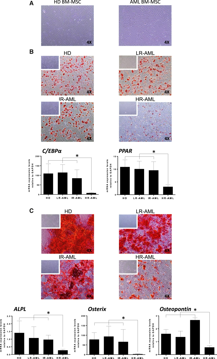
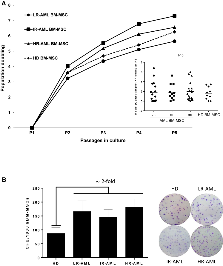
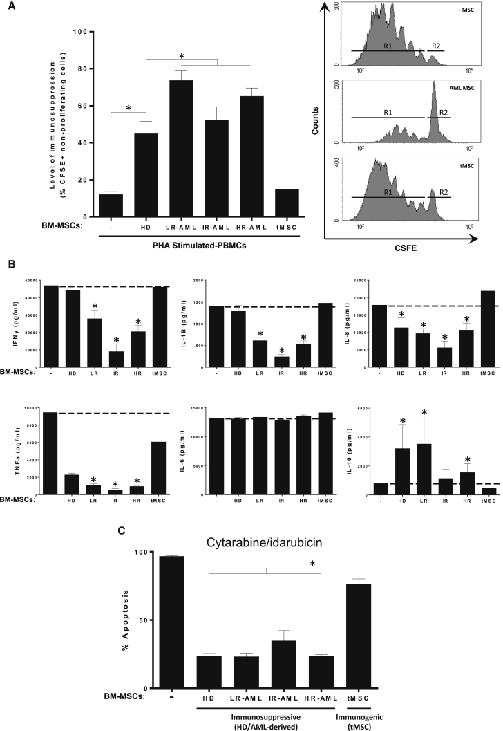
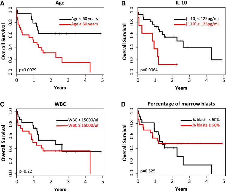
Bone marrow mesenchymal stem/stromal cells (BM-MSCs) are key components of the hematopoietic niche thought to have a direct role in leukemia pathogenesis. BM-MSCs from patients with acute myeloid leukemia (AML) have been poorly characterized due to disease heterogeneity. We report a functional, genetic, and immunological characterization of BM-MSC cultures from 46 AML patients, stratified by molecular/cytogenetics into low-risk (LR), intermediate-risk (IR), and high-risk (HR) subgroups. Stable MSC cultures were successfully established and characterized from 40 of 46 AML patients irrespective of the risk subgroup. AML-derived BM-MSCs never harbored tumor-specific cytogenetic/molecular alterations present in blasts, but displayed higher clonogenic potential than healthy donor (HD)-derived BM-MSCs. Although HD- and AML-derived BM-MSCs equally provided chemoprotection to AML cells in vitro, AML-derived BM-MSCs were more immunosuppressive/anti-inflammatory, enhanced suppression of lymphocyte proliferation, and diminished secretion of pro-inflammatory cytokines. Multivariate analysis revealed that the level of interleukin-10 produced by AML-derived BM-MSCs as an independent prognostic factor negatively affected overall survival. Collectively our data show that AML-derived BM-MSCs are not tumor related, but display functional differences contributing to therapy resistance and disease evolution.
Keywords: AML; BM-MSC; IL-10; characterization; chemoprotection; immunosuppression; risk-stratification.
Copyright © 2017 The Authors. Published by Elsevier Inc. All rights reserved.
Figures
References
-
- Arber D.A., Orazi A., Hasserjian R., Thiele J., Borowitz M.J., Le Beau M.M., Bloomfield C.D., Cazzola M., Vardiman J.W. The 2016 revision to the World Health Organization classification of myeloid neoplasms and acute leukemia. Blood. 2016;127:2391–2405. - PubMed
-
- Bene M.C., Grimwade D., Haferlach C., Haferlach T., Zini G., European L. Leukemia diagnosis: today and tomorrow. Eur. J. Haematol. 2015;95:365–373. - PubMed
-
- Bennett J.M., Catovsky D., Daniel M.T., Flandrin G., Galton D.A., Gralnick H.R., Sultan C. Proposed revised criteria for the classification of acute myeloid leukemia. A report of the French-American-British Cooperative Group. Ann. Intern. Med. 1985;103:620–625. - PubMed
-
- Bernardo M.E., Fibbe W.E. Mesenchymal stromal cells: sensors and switchers of inflammation. Cell Stem Cell. 2013;13:392–402. - PubMed
-
- Blau O., Hofmann W.K., Baldus C.D., Thiel G., Serbent V., Schumann E., Thiel E., Blau I.W. Chromosomal aberrations in bone marrow mesenchymal stroma cells from patients with myelodysplastic syndrome and acute myeloblastic leukemia. Exp. Hematol. 2007;35:221–229. - PubMed
MeSH terms
Substances
LinkOut - more resources
Full Text Sources
Other Literature Sources
Medical
Research Materials
Miscellaneous
