. 2017 Jul 5:276:85-91.
doi: 10.1016/j.toxlet.2017.05.015. Epub 2017 May 18.

Induction of proteotoxic stress by the mycotoxin patulin

Affiliations

Induction of proteotoxic stress by the mycotoxin patulin

Angel Guerra-Moreno et al. Toxicol Lett. .

Abstract

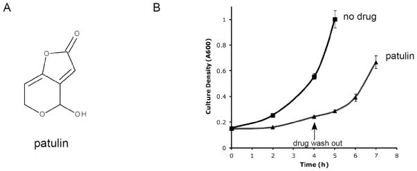
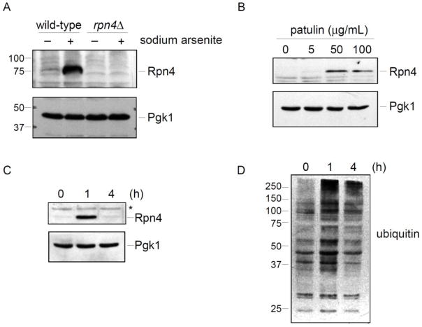
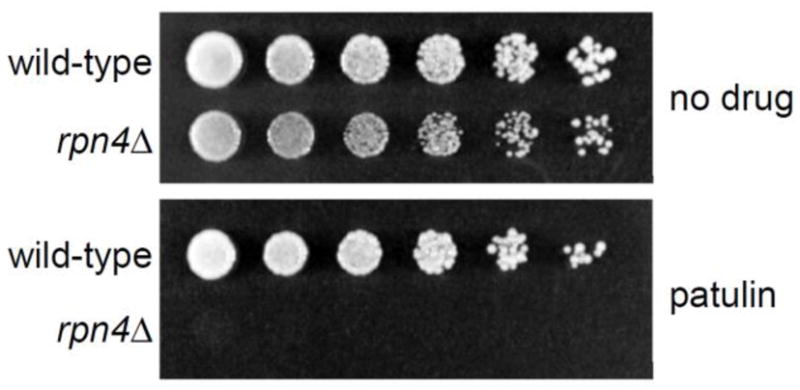
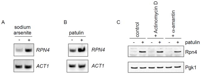
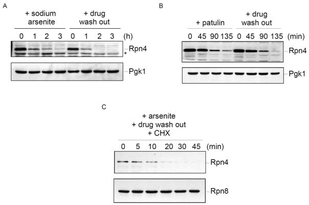
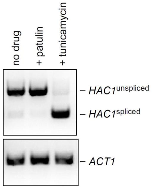
Patulin is a naturally occurring mycotoxin produced by a number of molds and may contaminate a wide variety of food products. In practice, patulin's main societal relevance concerns apple juice and its products. Multiple advisory bodies, including the U.S. Food and Drug Administration and the World Health Organization, recommend that producers monitor and limit patulin levels in apple juice products. The mechanism of patulin toxicity remains largely unknown. Here we show that patulin induces proteotoxic stress in the yeast S. cerevisiae. The transcription factor Rpn4 controls the abundance of the proteasome, the complex multisubunit protease that destroys proteins, including misfolded proteins. Rpn4 protein is strongly induced by patulin, and Rpn4 levels normalize over time, consistent with homeostatic regulation. A rpn4Δ mutant is highly sensitive to patulin, confirming the physiologic relevance of this response. Rpn4 is known to be regulated both transcriptionally and post-translationally. Patulin induces both pathways of regulation, but the post-transcriptional pathway predominates in controlling Rpn4 protein levels. These results indicate that proteotoxicity represents a major aspect of patulin toxicity. They not only have implications for patulin detoxification but in addition suggest the possibility of some potentially useful patulin applications.

Keywords: Patulin; Proteasome; Proteotoxicity; Rpn4; Yeast.


Figures

Figure 1
Figure 1. Patulin structure and reversible toxicity
A) Chemical structure of patulin. B) Growth of wild-type cells in liquid culture during treatment with patulin (50 μg/mL). After 4 hours of treatment, the drug was washed out. Error bars represent standard deviations from two independent cultures.
Figure 2
Figure 2. Patulin induces the Rpn4 stress response
A) Validation of the Rpn4 antibody. Wild-type and rpn4Δ strains were treated with sodium arsenite (1 mM) for one hour. Whole cell extracts were prepared and evaluated by SDS-PAGE followed by immunoblot with anti-Rpn4 antibody (upper panel) or anti-Pgk1 antibody (lower panel; loading control). B) Rpn4 protein levels after patulin treatment for one hour at the indicated concentrations. Whole cell extracts from a wild-type strain were prepared and analyzed by SDS-PAGE followed by immunoblot with anti-Rpn4 antibody (upper panel) or anti-Pgk1 antibody (lower panel; loading control). C) Temporal dynamics of the Rpn4 response after induction by patulin (50 μg/mL). Whole cell extracts from a wild-type strain were prepared and analyzed by SDS-PAGE followed by immunoblot with anti-Rpn4 antibody (upper panel) or anti-Pgk1 antibody (lower panel; loading control). Asterisk, non-specific immunoreactive band. D) Levels of ubiquitin conjugates in whole cell extracts, as determined by SDS-PAGE followed by immunoblot with anti-ubiquitin antibody. The same whole cell extracts were used to prepare the immunoblots in panels C and D. Therefore, the Pgk1 loading control of panel C applies to panel D as well.
Figure 3
Figure 3. Impaired growth of the rpn4Δ mutant after patulin treatment
Wild-type and rpn4Δ cells were spotted in three-fold serial dilutions on plates lacking or containing patulin (5 μg/mL) and cultured at 37°C for 4 days.
Figure 4
Figure 4. Induction of Rpn4 protein levels by patulin appears largely post-transcriptional
A–B) Induction of RPN4 transcription, as determined by RT-PCR, after treatment with sodium arsenite (1 mM) or patulin (50 μg/mL) for one hour. ACT1 serves as a control. C) Rpn4 protein levels after treatment with patulin (30 μg/mL) in the absence or presence of the transcriptional inhibitors actinomycin D (20 μg/mL) or α-amanitin (20 μg/mL). Whole cell extracts from a wild-type strain were prepared and analyzed by SDS-PAGE followed by immunoblot with anti-Rpn4 antibody (upper panel) or anti-Pgk1 antibody (lower panel; loading control).
Figure 5
Figure 5. Precise regulation of Rpn4 protein levels in response to stress
A) Steady-state Rpn4 protein levels after treatment with sodium arsenite (1 mM). The zero time point reflects Rpn4 induction after one hour of drug treatment. Sodium arsenite was then maintained in the culture or washed out, and Rpn4 levels were followed over time. Whole cell extracts were prepared and analyzed by SDS-PAGE followed by immunoblot with anti-Rpn4 antibody (upper panel) or anti-Pgk1 antibody (lower panel; loading control). B) Steady-state Rpn4 protein levels after treatment with patulin (50 μg/mL). The zero time point reflects Rpn4 induction after one hour of drug treatment. Extracts were prepared and analyzed as in panel A. C) Cycloheximide chase analysis of Rpn4 turnover. Rpn4 protein was induced by treatment with sodium arsenite (1 mM) for one hour. The drug was then washed out and cycloheximide (100 μg/mL) was added to inhibit new protein synthesis. Whole cell extracts were prepared and analyzed by SDS-PAGE followed by immunoblot with anti-Rpn4 antibody (upper panel) or anti-Rpn8 antibody (lower panel; loading control).
Figure 6
Figure 6. Patulin fails to induce the unfolded protein response
HAC1 splicing, which is a key aspect of the unfolded protein response, was measured by RT-PCR after treatment with patulin (30 μg/mL) for one hour. Tunicamycin, a known inducer of the unfolded protein response, was used as a positive control. ACT1 (lower panel) serves as a control.

Similar articles

Cited by

References

    1. Bennett JW, Klich M. Mycotoxins. Clin Microbiol Rev. 2003;16:497–516. - PMC - PubMed
    1. Bensaude O. Inhibiting eukaryotic transcription: Which compound to choose? How to evaluate its activity? Transcription. 2011;2:103–108. - PMC - PubMed
    1. Cox JS, Walter P. A novel mechanism for regulating activity of a transcription factor that controls the unfolded protein response. Cell. 1996;87:391–404. - PubMed
    1. Finley D, Ulrich HD, Sommer T, Kaiser P. The ubiquitin-proteasome system of Saccharomyces cerevisiae. Genetics. 2012;192:319–60. - PMC - PubMed
    1. Fliege R, Metzler M. The mycotoxin patulin induces intra- and intermolecular protein crosslinks in vitro involving cysteine, lysine, and histidine side chains, and alpha-amino groups. Chem Biol Interact. 1999;123:85–103. - PubMed

MeSH terms

LinkOut - more resources