

Genetic deletion of Sost or pharmacological inhibition of sclerostin prevent multiple myeloma-induced bone disease without affecting tumor growth
- PMID: 28529307
- PMCID: PMC5699973
- DOI: 10.1038/leu.2017.152
Genetic deletion of Sost or pharmacological inhibition of sclerostin prevent multiple myeloma-induced bone disease without affecting tumor growth
Abstract
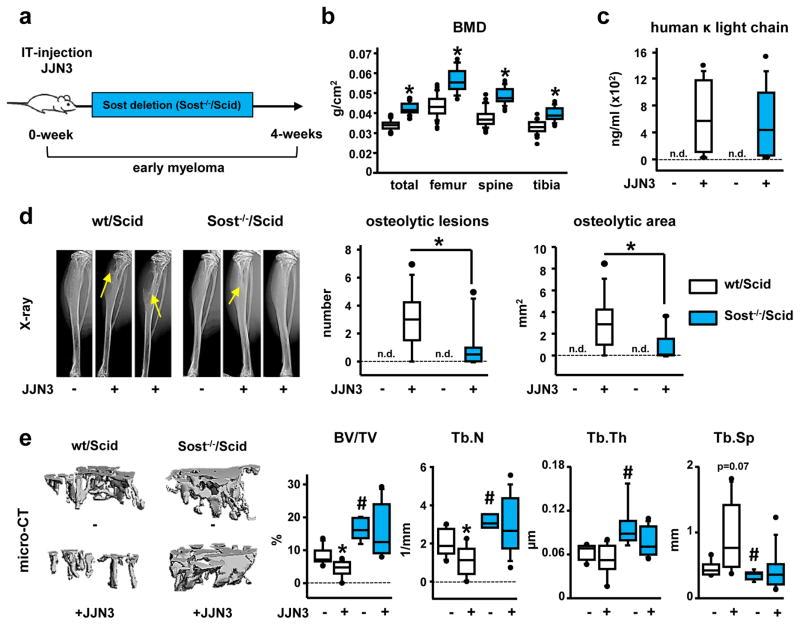
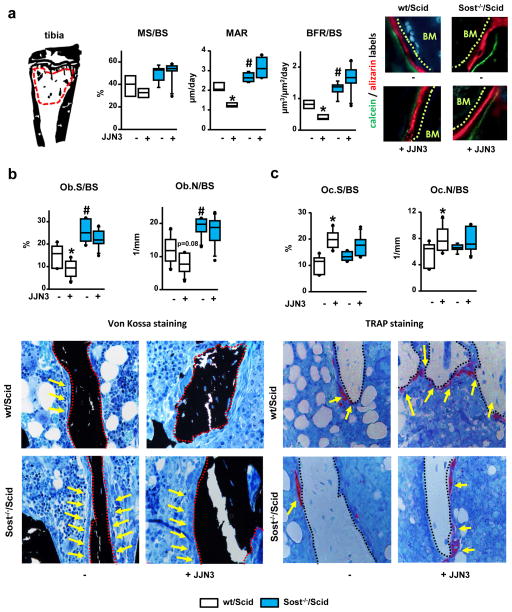
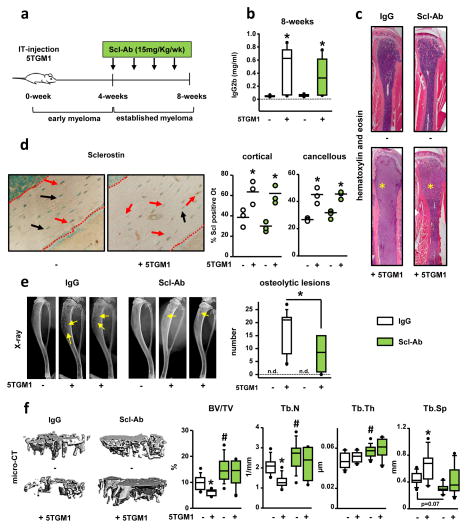
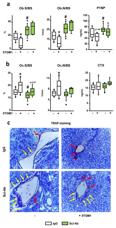
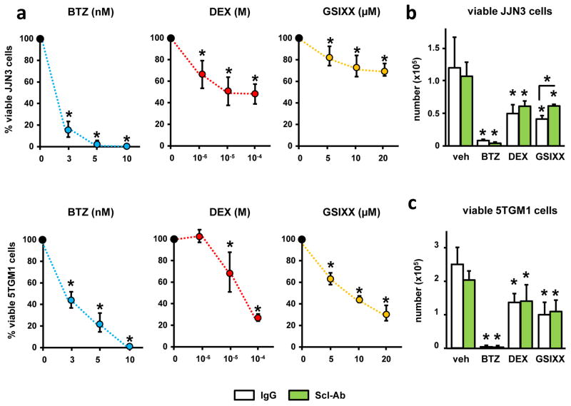
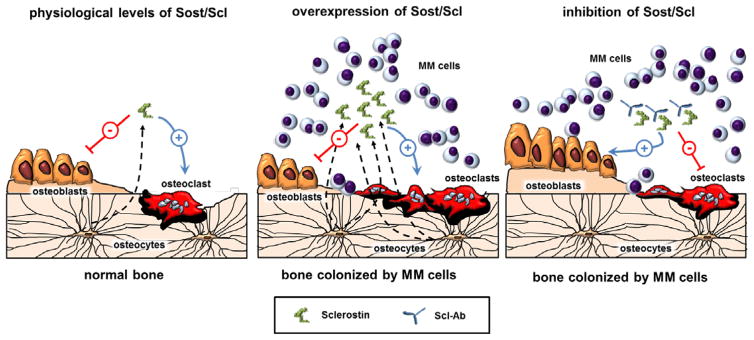
Multiple myeloma (MM) causes lytic bone lesions due to increased bone resorption and concomitant marked suppression of bone formation. Sclerostin (Scl), an osteocyte-derived inhibitor of Wnt/β-catenin signaling, is elevated in MM patient sera and increased in osteocytes in MM-bearing mice. We show here that genetic deletion of Sost, the gene encoding Scl, prevented MM-induced bone disease in an immune-deficient mouse model of early MM, and that administration of anti-Scl antibody (Scl-Ab) increased bone mass and decreases osteolysis in immune-competent mice with established MM. Sost/Scl inhibition increased osteoblast numbers, stimulated new bone formation and decreased osteoclast number in MM-colonized bone. Further, Sost/Scl inhibition did not affect tumor growth in vivo or anti-myeloma drug efficacy in vitro. These results identify the osteocyte as a major contributor to the deleterious effects of MM in bone and osteocyte-derived Scl as a promising target for the treatment of established MM-induced bone disease. Further, Scl did not interfere with efficacy of chemotherapy for MM, suggesting that combined treatment with anti-myeloma drugs and Scl-Ab should effectively control MM growth and bone disease, providing new avenues to effectively control MM and bone disease in patients with active MM.
Conflict of interest statement
G.D.R. has received consulting honoraria from Amgen Inc. S.A.K is an employee of Lilly Research Laboratories. The remaining authors declare no competing financial interests.
G.D.R. has received consulting honoraria from Amgen Inc. S.A.K is an employee of Lilly Research Laboratories. The remaining authors declare no competing financial interests.
Figures
Similar articles
-
Role of Osteocytes in Myeloma Bone Disease: Anti-sclerostin Antibody as New Therapeutic Strategy.Front Immunol. 2018 Oct 24;9:2467. doi: 10.3389/fimmu.2018.02467. eCollection 2018. Front Immunol. 2018. PMID: 30410490 Free PMC article. Review.
-
Bidirectional Notch Signaling and Osteocyte-Derived Factors in the Bone Marrow Microenvironment Promote Tumor Cell Proliferation and Bone Destruction in Multiple Myeloma.Cancer Res. 2016 Mar 1;76(5):1089-100. doi: 10.1158/0008-5472.CAN-15-1703. Epub 2016 Feb 1. Cancer Res. 2016. PMID: 26833121 Free PMC article.
-
Inhibiting the osteocyte-specific protein sclerostin increases bone mass and fracture resistance in multiple myeloma.Blood. 2017 Jun 29;129(26):3452-3464. doi: 10.1182/blood-2017-03-773341. Epub 2017 May 17. Blood. 2017. PMID: 28515094 Free PMC article.
-
Regulation of Sclerostin Expression in Multiple Myeloma by Dkk-1: A Potential Therapeutic Strategy for Myeloma Bone Disease.J Bone Miner Res. 2016 Jun;31(6):1225-34. doi: 10.1002/jbmr.2789. Epub 2016 Feb 19. J Bone Miner Res. 2016. PMID: 26763740 Free PMC article.
-
The effects of bortezomib on bone disease in patients with multiple myeloma.Cancer. 2014 Mar 1;120(5):618-23. doi: 10.1002/cncr.28481. Epub 2013 Nov 18. Cancer. 2014. PMID: 24249482 Review.
Cited by
-
Development and characterization of three cell culture systems to investigate the relationship between primary bone marrow adipocytes and myeloma cells.Front Oncol. 2023 Jan 11;12:912834. doi: 10.3389/fonc.2022.912834. eCollection 2022. Front Oncol. 2023. PMID: 36713534 Free PMC article.
-
Sclerostin inhibition alleviates breast cancer-induced bone metastases and muscle weakness.JCI Insight. 2019 Apr 9;5(9):e125543. doi: 10.1172/jci.insight.125543. JCI Insight. 2019. PMID: 30965315 Free PMC article.
-
The multifunctional role of Notch signaling in multiple myeloma.J Cancer Metastasis Treat. 2021;7:20. doi: 10.20517/2394-4722.2021.35. Epub 2021 Apr 14. J Cancer Metastasis Treat. 2021. PMID: 34778567 Free PMC article.
-
Multiple Myeloma and Bone: The Fatal Interaction.Cold Spring Harb Perspect Med. 2018 Aug 1;8(8):a031286. doi: 10.1101/cshperspect.a031286. Cold Spring Harb Perspect Med. 2018. PMID: 29229668 Free PMC article. Review.
-
Targeting Notch Inhibitors to the Myeloma Bone Marrow Niche Decreases Tumor Growth and Bone Destruction without Gut Toxicity.Cancer Res. 2021 Oct 1;81(19):5102-5114. doi: 10.1158/0008-5472.CAN-21-0524. Epub 2021 Aug 4. Cancer Res. 2021. PMID: 34348968 Free PMC article.
References
-
- Roodman GD. Pathogenesis of myeloma bone disease. J Cell Biochem. 2010;109:283–291. - PubMed
-
- Silbermann R, Roodman GD. Bone effects of cancer therapies: pros and cons. Curr Opin Support Palliat Care. 2011;5:251–257. - PubMed
-
- Roodman GD. Targeting the bone microenvironment in multiple myeloma. J Bone Miner Metab. 2010;28:244–250. - PubMed
-
- Roodman GD. Role of the bone marrow microenvironment in multiple myeloma. J Bone Miner Res. 2002;17:1921–1925. - PubMed
MeSH terms
Substances
Grants and funding
LinkOut - more resources
Full Text Sources
Other Literature Sources
Medical
Molecular Biology Databases
Research Materials
Miscellaneous
