. 2017 Mar 11;13(4):413-425.
doi: 10.7150/ijbs.17508. eCollection 2017.

MiR-696 Regulates C2C12 Cell Proliferation and Differentiation by Targeting CNTFRα

Affiliations

MiR-696 Regulates C2C12 Cell Proliferation and Differentiation by Targeting CNTFRα

Han Wang et al. Int J Biol Sci. .

Abstract

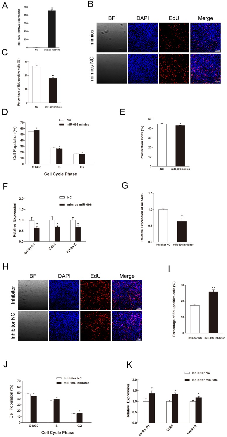
Micro-696 (miR-696) has been previously known as an exercise related miRNA, which has a profound role in fatty acid oxidation and mitochondrial biogenesis of skeletal muscle. However, its role in skeletal myoblast proliferation and differentiation is still unclear. In this study, we found that miR-696 expressed highly in skeletal muscle and reduced during C2C12 myoblasts differentiation. MiR-696 overexpression repressed C2C12 myoblast proliferation and myofiber formation, while knockdown of endogenous miR-696 expression showed opposite results. During myogenesis, we observed an inversed expression pattern between miR-696 and CNTFRα in vitro, and demonstrated that miR-696 could specifically target CNTFRα and repress the expression of CNTFRα. Additionally, we further found that knockdown of CNTFRα suppressed the proliferation and differentiation of C2C12 cells. Taking all things together, we propose a novel insight that miR-696 down-regulates C2C12 cell myogenesis by inhibiting CNTFRα expression.

Keywords: CNTFRα; MiR-696; myoblast proliferation and differentiation..


Conflict of interest statement

Competing Interests: The authors have declared that no competing interest exists.

Figures

Figure 1
Figure 1
Tissue distribution of miR-696 and its expression pattern during C2C12 myoblast differentiation. (A) Relative expression of miR-696 in different mouse tissues. (B) Expression of miR-696 in mouse EDL and SOL muscles. (C) Morphological images of C2C12 myoblasts cultured in GM or in DM for 2, 4 and 6 days. (D) The mRNA level of MyHC and MyoG in 0, 2, 4, and 6 days of cell differentiation. The fold change was relative to day 0 of GM expression. GAPDH was used as the reference gene for Q-PCR. (E) The protein level of MyHC and MyoG in 0, 2, 4, and 6 days of cell differentiation. The fold change was relative to day 0 of GM expression.β-actin as controls for western blotting. (F) Expression of miR-696 during proliferation. The fold change was relative to 30% cell confluence. (G). Expression of miR-696 in C2C12 cells differentiated for 0, 2, 4, and 6 days. The fold change was relative to day 0 of GM expression. U6 was used as the reference gene. Results are expressed as mean ± S.E.M. (n = 3). *P<0.05; **P<0.01.
Figure 2
Figure 2
MiR-696 represses the proliferation of C2C12 cells. (A) The expression of miR-696 was detected using qPCR in myoblast transfected with miR-696 mimics or NC. (B) After transfection with miR-696 mimics or NC for 24 h, cells were fixed for EdU (red). The “large things” in Fig 2B was not a cell but some water-drops. It might because of the lid wasn't on tight caused by our carelessness. Scale bar = 100 μm. (C) The proportion of EdU-positive cells were presented. (D) C2C12 cells were collected for PI flow cytometry. (E) The proliferation index was calculated. (F) Expression of cell cycle related genes at 24 h post-transfection. (G) miR-696 expression was detected in myoblasts after transfected with miR-696 inhibitor or inhibitor NC. (H) After transfection with miR-696 inhibitor or inhibitor NC for 24 h, proliferating C2C12 cells were fixed for EdU (red). Scale bar = 100 μm. (I) The proportion of EdU-positive cells were compared between miR-696 inhibitor group and inhibitor-NC group. (J) Cell cycle distribution was detected by PI flow cytometry. (K) Expression of cell cycle related genes at 24 h post-transfection. Results are expressed as mean ± S.E.M. (n = 3). *P<0.05; **P<0.01.
Figure 3
Figure 3
MiR-696 inhibits myogenic differentiation of C2C12 cells. (A) The expression of miR-696 were detected by qPCR in myoblast transfected with miR-696 mimics or NC duplexes at day 3 of differentiation medium (DM). (B) After transfection with miR-696 mimics or NC at GM and collected at day 3 of DM, the MyHC and MyoG mRNA expression were determined. (C) The MyHC and MyoG protein expression were determined at 3 days of DM. (D) MyHC (green) was detected with Immunofluorescence after transfection with miR-696 mimics or NC duplexes at day 3 of DM. Scale bar = 50 μm. (E) The expression of miR-696 were detected in myoblast after transfected with inhibitor miR-696 or inhibitor NC at day 3s of differentiation medium (DM). (F) After transfection with inhibitor miR-696 or inhibitor NC at GM and collected at day 3 of DM, MyHC and MyoG mRNA expression were determined. (G) The MyHC and MyoG protein expression were determined at day 3 of DM. (H) MyHC (green) was detected with Immunofluorescence after day 3 of miR-696 inhibitor or inhibitor NC transfection at DM. Scale bar = 50 μm. Results are expressed as mean ± S.E.M. (n = 3). *P<0.05; **P<0.01.
Figure 4
Figure 4
CNTFRα is a direct target of miR-696. (A) Sequence of miR-696 and its predicted binding region in CNTFRα 3'UTR (red). (B) The sketch map of the dual-luciferase reporter vector pmirGLO. The putative miR-696 target site of the CNTFRα 3'UTRs and mutation target site were inserted into the 3' end of the firefly luciferase gene (luc2). The expression of the Renilla luciferase (hRluc-neo) was treated as an internal normalization control. (C) C2C12 and HEK293T cells transfected with miR-696 mimics or NC were co-transfected with the pCNTFRα vector or mutant dual-luciferase vector. The relative luciferase activity was assayed 24 h later. (D) The mRNA expression of CNTFRα in C2C12 cells during proliferation. (E) The protein expression of CNTFRα in C2C12 cells during proliferation. (F) The mRNA expression of CNTFRα in 0, 2, 4, and 6 days of cell differentiation. The fold change was relative to day 0 of GM expression. GAPDH was used as the reference gene in qPCR. (G) The protein expression of CNTFRα in in 0, 2, 4, and 6 days of cell differentiation. β-actin was treated as control protein in western blotting assay. (H) After transfection for 24 h with miR-696 mimics, inhibitor miR-696, NC, or inhibitor NC for 24h, the mRNA expression of CNTFRα was detected by qPCR in proliferating C2C12 cells. (I) The protein expression of CNTFRα after transfection for 24 h with miR-696 mimics, inhibitor miR-696, NC, or inhibitor NC for 24h was detected by western blot in proliferating C2C12 cells. (J) The mRNA level of CNTFRα was determined by qPCR in myoblast transfected with miR-696 mimics, inhibitor miR-696, NC, or inhibitor NC at day 3 of differentiation medium (DM). (K) The protein level of CNTFRα in myoblast transfected with miR-696 mimics, inhibitor miR-696, NC, or inhibitor NC at day 3 of differentiation medium (DM). Results are expressed as mean ± S.E.M. (n = 3). *P<0.05; **P<0.01.
Figure 5
Figure 5
Down-regulation of CNTFRα inhibited myoblast proliferation and differentiation. (A) The expression of CNTFRα mRNA was detected at 24 h post-transfection in GM. (B) The expression of CNTFRα protein was detected at 24 h post-transfection in GM. (C) After transfection with siCNTFRα or NC for 24h, cells were fixed for EdU (red). Scale bar = 100 μm. The proportion of EdU-positive cells were presented. (D) The cell cycle distribution was detected by PI flow cytometry. The proliferation index was calculated. (E) Expression of cell cycle related genes at 24 h post-transfection. (F) The mRNA level of CNTFRα, MyHC and MyoG were detected by qPCR in myoblast transfected with siCNTFRα or NC at day 3 of differentiation medium (DM). (G) The protein level of CNTFRα, MyHC and MyoG were detected by western blotting in myoblast transfected with siCNTFRα or NC at day 3 of differentiation medium (DM). (H) MyHC (green) was detected with Immunofluorescence after 3 days of siCNTFRα or NC transfection at DM. Scale bar = 50 μm. Results are expressed as mean ± S.E.M. (n = 3). *P<0.05; **P<0.01.

Similar articles

Cited by

References

    1. Buckingham M, Bajard L, Chang T, Daubas P, Hadchouel J, Meilhac S. et al. The formation of skeletal muscle: from somite to limb. Journal of anatomy. 2003;202:59–68. - PMC - PubMed
    1. Perry RL, Rudnick MA. Molecular mechanisms regulating myogenic determination and differentiation. Frontiers in bioscience: a journal and virtual library. 2000;5:D750–67. - PubMed
    1. Buckingham M, Rigby PW. Gene regulatory networks and transcriptional mechanisms that control myogenesis. Developmental cell. 2014;28:225–38. - PubMed
    1. Bartel DP. MicroRNAs: target recognition and regulatory functions. Cell. 2009;136:215–33. - PMC - PubMed
    1. Hutvagner G, Zamore PD. A microRNA in a multiple-turnover RNAi enzyme complex. Science. 2002;297:2056–60. - PubMed

Publication types

MeSH terms

Substances

LinkOut - more resources