

The role of germ cell loss during primordial follicle assembly: a review of current advances
- PMID: 28529453
- PMCID: PMC5436565
- DOI: 10.7150/ijbs.18836
The role of germ cell loss during primordial follicle assembly: a review of current advances
Abstract
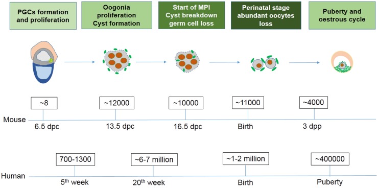
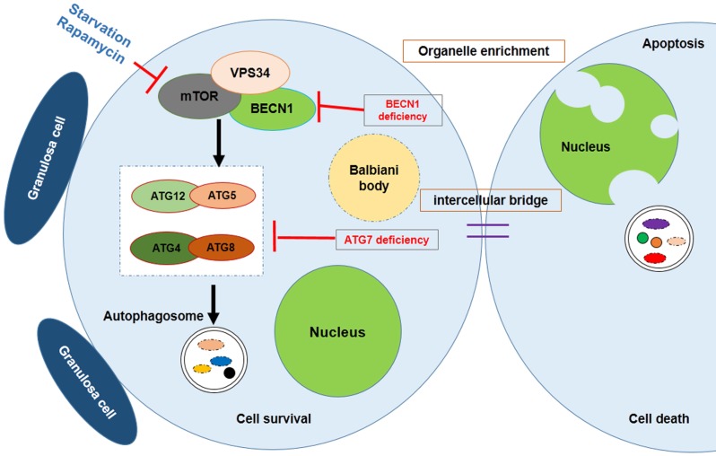
In most female mammals, early germline development begins with the appearance of primordial germ cells (PGCs), and develops to form mature oocytes following several vital processes. It remains well accepted that significant germ cell apoptosis and oocyte loss takes place around the time of birth. The transition of the ovarian environment from fetal to neonatal, coincides with the loss of germ cells and the timing of follicle formation. All told it is common to lose approximately two thirds of germ cells during this transition period. The current consensus is that germ cell loss can be attributed, at least in part, to programmed cell death (PCD). Recently, autophagy has been implicated as playing a part in germ cell loss during the time of parturition. In this review, we discuss the major opinions and mechanisms of mammalian ovarian PCD during the process of germ cell loss. We also pay close attention to the function of autophagy in germ cell loss, and speculate that autophagy may also serve as a critical and necessary process during the establishment of primordial follicle pool.
Keywords: Apoptosis; Autophagy; Germ cell cyst; Germ cell loss; Primordial follicle assembly.
Conflict of interest statement
Competing Interests: The authors have declared that no competing interest exists.
Figures
References
-
- Borum K. Oogenesis in the mouse. A study of the meiotic prophase. Exp Cell Res. 1961;24:495–507. - PubMed
-
- McClellan KA, Gosden R, Taketo T. Continuous loss of oocytes throughout meiotic prophase in the normal mouse ovary. Dev Biol. 2003;258:334–348. - PubMed
-
- Morita Y, Tilly JL. Oocyte apoptosis: like sand through an hourglass. Dev Biol. 1999;213:1–17. - PubMed
-
- Pepling ME, Spradling AC. Mouse ovarian germ cell cysts undergo programmed breakdown to form primordial follicles. Dev Biol. 2001;234:339–351. - PubMed
-
- Baker TG. Oogenesis and ovarian development. In: Balin H, Glasser S, editors. Reproductive Biology. Amsterdam: Excerpta Medica; 1972.
Publication types
MeSH terms
LinkOut - more resources
Full Text Sources
Other Literature Sources
