Review
. 2017 Apr 7;7(6):1543-1588.
doi: 10.7150/thno.15625. eCollection 2017.

Role of the CXCL8-CXCR1/2 Axis in Cancer and Inflammatory Diseases

Affiliations
Review

Role of the CXCL8-CXCR1/2 Axis in Cancer and Inflammatory Diseases

Helen Ha et al. Theranostics. .

Abstract

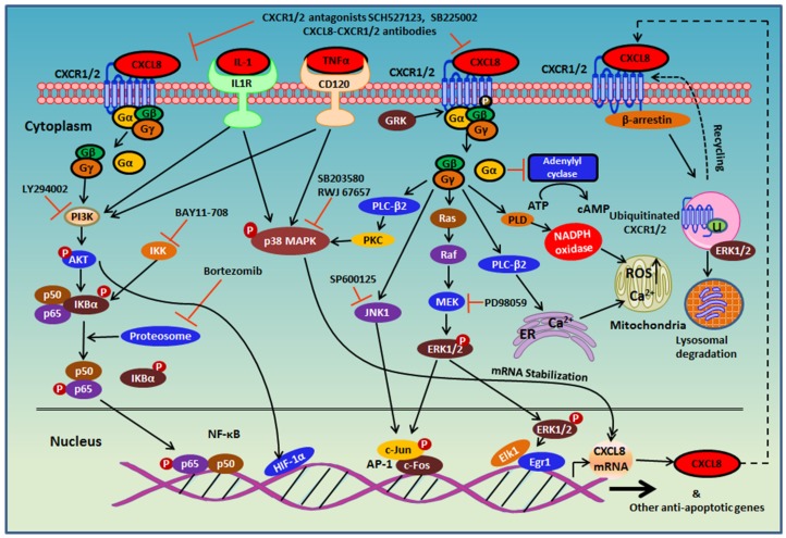
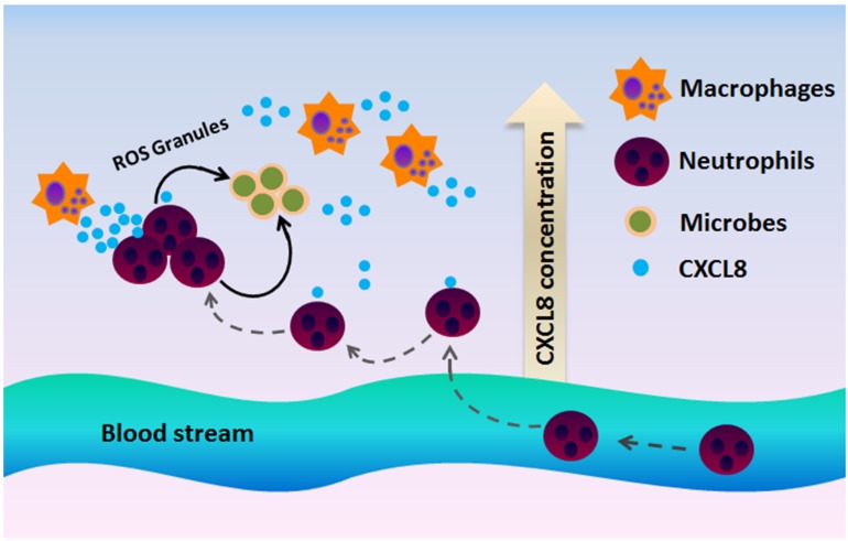
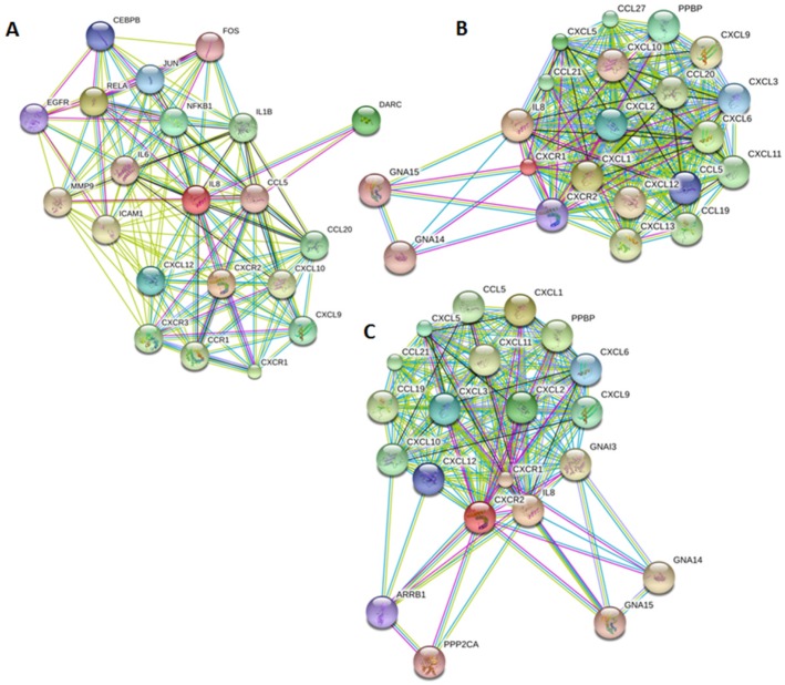
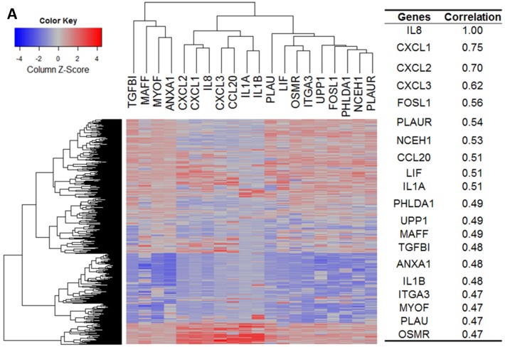
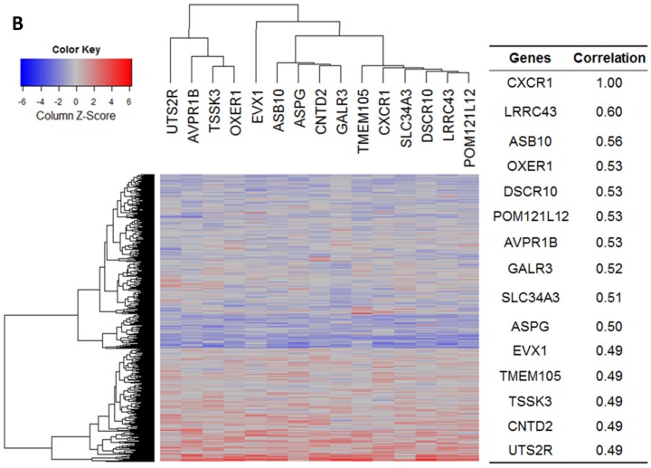
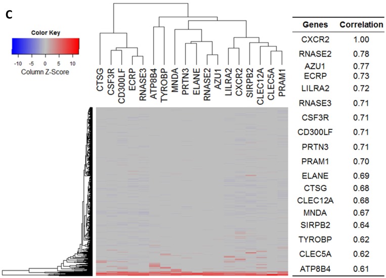
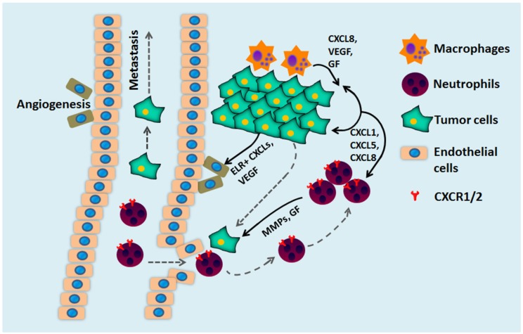
The chemokine receptors CXCR1/2 and their ligand CXCL8 are essential for the activation and trafficking of inflammatory mediators as well as tumor progression and metastasis. The CXCL8-CXCR1/2 signaling axis is involved in the pathogenesis of several diseases including chronic obstructive pulmonary diseases (COPD), asthma, cystic fibrosis and cancer. Interaction between CXCL8 secreted by select cancer cells and CXCR1/2 in the tumor microenvironment is critical for cancer progression and metastasis. The CXCL8-CXCR1/2 axis may play an important role in tumor progression and metastasis by regulating cancer stem cell (CSC) proliferation and self-renewal. During the past two decades, several small-molecule CXCR1/2 inhibitors, CXCL8 releasing inhibitors, and neutralizing antibodies against CXCL8 and CXCR1/2 have been reported. As single agents, such inhibitors are expected to be efficacious in various inflammatory diseases. Several preclinical studies suggest that combination of CXCR1/2 inhibitors along with other targeted therapies, chemotherapies, and immunotherapy may be effective in treating select cancers. Currently, several of these inhibitors are in advanced clinical trials for COPD, asthma, and metastatic breast cancer. In this review, we provide a comprehensive analysis of the role of the CXCL8-CXCR1/2 axis and select genes co-expressed in this pathway in disease progression. We also discuss the latest progress in developing small-molecule drugs targeting this pathway.

Keywords: CXCL8; CXCR1; CXCR2; antibody.; cancer; cancer stem cells; chronic obstructive pulmonary diseases; inhibitor; tumor microenvironment.


Conflict of interest statement

Competing Interests: The authors have declared that no competing interest exists.

Figures

Figure 1
Figure 1
Phylogenetic tree of chemokine receptors. The phylogenetic tree was generated from sequence homology using the tools in the GPCR database (gpcrdb.org).
Figure 2
Figure 2
Structures of CXCL8 and its receptors CXCR1/2. A. CXCL8 protein structure: The dimer structure was generated from the Protein Data Bank code PDB ID 1ICW. Two monomers are colored as green and cyan. The ELR motif is colored as yellow and CXC motif is in blue. B. CXCR2 2D structural domains: The N-terminus (extracellular face) of CXCR2 is critical for ligand binding and specificity. Transmembrane domain 4 and extracellular loop 2 is also important for ligand binding. The G-protein couples to the C-terminus (cytoplasmic face) of CXCR2 and involves intracellular loop 3. Several proteins, such as G protein coupled receptor kinase 6 (GRK6), vasodilator-stimulated phosphoprotein (VASP), β-arrestin1/2, adaptor protein-2 (AP-2), protein phosphatase 2A (PP2A) also associate with the C-terminus of CXCR2 to mediate different signaling cascades. C. CXCR1/2 3D structure: CXCR1 solid state NMR structure (PDB: 2LNL, green) aligned with CXCR2 homology model (blue) . The predicted binding mode of the CXCR2 antagonist SCH527123 (brown) in the allosteric site located inside the CXCR2 receptor .
Figure 3
Figure 3
CXCL8-CXCR1/2 signaling cascades and receptor recycling. CXCL8 binding to CXCR1/2 activates several G-protein-mediated signaling cascades. Receptor activation immediately leads to the dissociation of the Gαi subunit from the βγ subunits, and subsequently activates growth and stress kinases such as ERK1/2, JNK1, and p38. G-protein activation also induces rapid intracellular Ca2+ mobilization released from the endoplasmic reticulum (ER) and inhibition of adenylyl cyclase resulting in decreased cyclic AMP production. CXCR1/2 activation also leads to receptor phosphorylation on the C-terminus by GRK2/6 and recruitment of β-arrestin1/2 to mediate receptor internalization. Internalized receptors are either recycled back to the cell surface or routed to lysosomes for degradation.
Figure 4
Figure 4
CXCR1 and CXCR2 mediate neutrophil recruitment during infection. In the presence of a microbial infection, macrophages at the site of infection begin to secrete CXCL8 to attract CXCR1/2-expressing neutrophils to the site of infection. Since CXCR2 is more sensitive to low ligand concentrations, CXCR2 is believed to play a more important role at recruiting neutrophils to the site of infection, whereas CXCR1 mediates oxidative burst and granule release to combat the microbes at the site of infection.
Figure 5
Figure 5
Genes implicated in the CXCL8-CXCR1/2 axis. Protein-protein interaction plots generated from STRING version 10 database using A. CXCL8 (IL8), B. CXCR1 and C. CXCR2 as queries. Pictures are top 20 genes connected to CXCL8 or CXCR1/2 either through physical interactions (experiments), co-expression, text mining, neighborhood on the genome, gene-fusion, database, co-occurrence and homology.
Figure 6
Figure 6
Gene expression heat map for CXCL8 (IL8), CXCR1/2 co-expressed genes from the CCLE as well as co-expression correlation. A. Gene expression heat map and correlations for CXCL8 (IL8) co-expressed genes. The top 20 genes are listed. B. Gene expression heat map and correlations for CXCR1 co-expressed genes. C. Gene expression heat map and correlations for CXCR2 co-expressed genes.
Figure 6
Figure 6
Gene expression heat map for CXCL8 (IL8), CXCR1/2 co-expressed genes from the CCLE as well as co-expression correlation. A. Gene expression heat map and correlations for CXCL8 (IL8) co-expressed genes. The top 20 genes are listed. B. Gene expression heat map and correlations for CXCR1 co-expressed genes. C. Gene expression heat map and correlations for CXCR2 co-expressed genes.
Figure 7
Figure 7
CXCL8, CXCR1/2 expression in cancer vs normal tissues from Oncomine analysis. Cancer vs normal tissue expression of CXCL8, CXCR1 and CXCR2 (A) where threshold p-value = 0.0001, fold change = 2 and gene rank = top 10%. CXCL8 expression (Log2 median) in patient samples of Kaiser Colon (B), Hu Esophagus (C), Ye Head-Neck (D) and Badea Pancreas (E). AC = Adenocarcinoma.
Figure 8
Figure 8
Gene expression data of CXCL8, CXCR1/2 in the panel of cancer cell lines. Gene expression of CXCL8, CXCR1/2 in the NCI-60 cell lines (A) and 1036 cell lines from the Cancer Cell Line Encyclopedia (CCLE) (B).
Figure 9
Figure 9
CXCL8 and CXCR1/2 gene expression distribution in normal tissues. Gene expression data of CXCL8 (A), CXCR1 (B) and CXCR2 (C) in various human tissues (NextBio, http://www.nextbio.com). Data are plotted as mean ± standard deviation. Dotted line represents the average value of all tissues.
Figure 9
Figure 9
CXCL8 and CXCR1/2 gene expression distribution in normal tissues. Gene expression data of CXCL8 (A), CXCR1 (B) and CXCR2 (C) in various human tissues (NextBio, http://www.nextbio.com). Data are plotted as mean ± standard deviation. Dotted line represents the average value of all tissues.
Figure 10
Figure 10
shRNA activity of CXCL8 (A), CXCR1 (B) and CXCR2 (C) in a panel of cancer cell lines. Data obtained from Project Achilles Data Portal of Broad Institute (https://www.broadinstitute.org/achilles ). shRNA score denotes log2 based decrease in CXCL8, CXCR1 or CXCR2 shRNA compared to pooled shRNA in cancer cell lines after several rounds of proliferation post-shRNA infection. A negative shRNA score indicates decreased cancer cell proliferation post-shRNA transfection.
Figure 11
Figure 11
The multiple roles of CXCL chemokines and CXCR1/2 during tumor development. CXCR1/2 and CXCL promote tumor growth through several mechanisms. Secretion of CXCL8 by tumor cells and tumor-associated macrophages stimulates cancer cell proliferation, survival, and chemoresistance. CXCL8 secretion also mediates neutrophil recruitment to the tumor site and stimulates neutrophils to secrete growth factors (GF) and matrix metalloproteinase (MMPs) to facilitate cancer cell migration, invasion, and metastases. CXCL8 stimulates CXCR2-expressing endothelial cells that form blood vessels within the tumor and stimulate tumor angiogenesis.

References

    1. Le Y, Zhou Y, Iribarren P, Wang J. Chemokines and chemokine receptors: their manifold roles in homeostasis and disease. Cell Mol Immunol. 2004;1:95–104. - PubMed
    1. Rollins BJ. Chemokines. Blood. 1997;90:909–28. - PubMed
    1. Debnath B, Xu S, Grande F, Garofalo A, Neamati N. Small molecule inhibitors of CXCR4. Theranostics. 2013;3:47–75. - PMC - PubMed
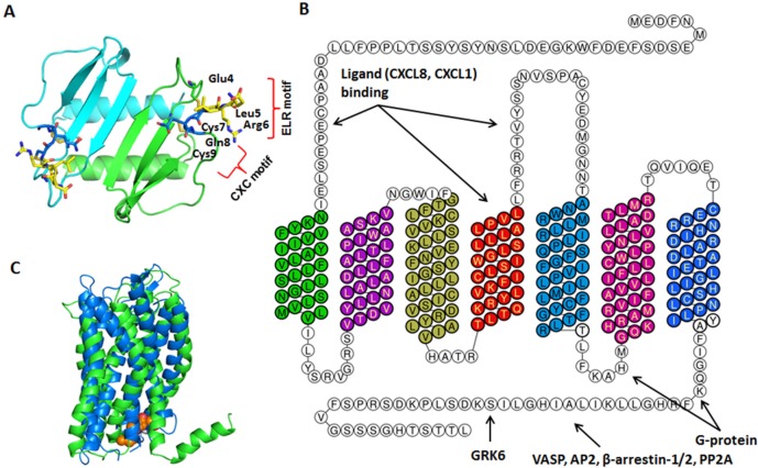
    1. Hebert CA, Vitangcol RV, Baker JB. Scanning mutagenesis of interleukin-8 identifies a cluster of residues required for receptor binding. J Biol Chem. 1991;266:18989–94. - PubMed
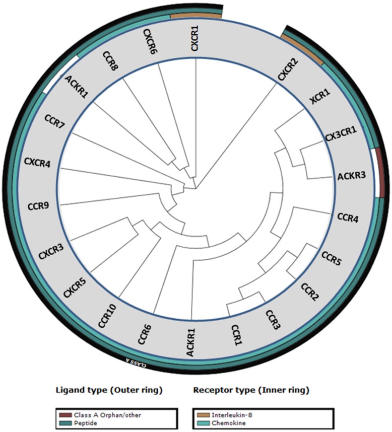
    1. GPCRdb. http://gpcrdb.org/

Publication types