

On-chip density-based purification of liposomes
- PMID: 28529672
- PMCID: PMC5422205
- DOI: 10.1063/1.4983174
On-chip density-based purification of liposomes
Abstract
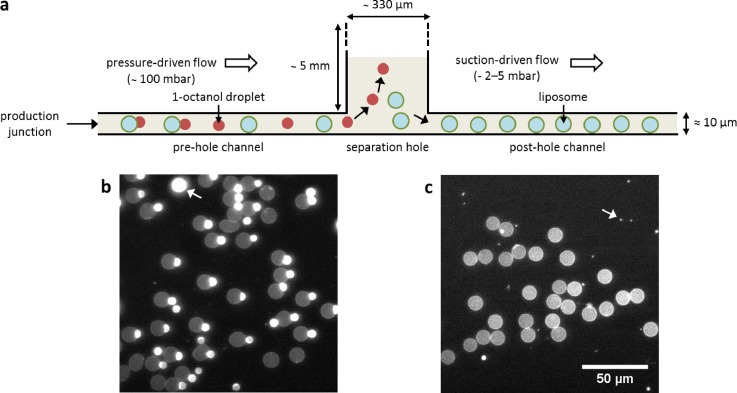
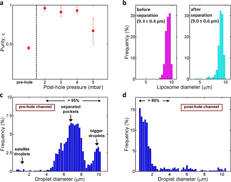
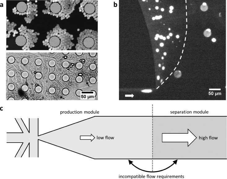
Due to their cell membrane-mimicking properties, liposomes have served as a versatile research tool in science, from membrane biophysics and drug delivery systems to bottom-up synthetic cells. We recently reported a novel microfluidic method, Octanol-assisted Liposome Assembly (OLA), to form cell-sized, monodisperse, unilamellar liposomes with excellent encapsulation efficiency. Although OLA provides crucial advantages over alternative methods, it suffers from the presence of 1-octanol droplets, an inevitable by-product of the production process. These droplets can adversely affect the system regarding liposome stability, channel clogging, and imaging quality. In this paper, we report a density-based technique to separate the liposomes from droplets, integrated on the same chip. We show that this method can yield highly pure (>95%) liposome samples. We also present data showing that a variety of other separation techniques (based on size or relative permittivity) were unsuccessful. Our density-based separation approach favourably decouples the production and separation module, thus allowing freshly prepared liposomes to be used for downstream on-chip experimentation. This simple separation technique will make OLA a more versatile and widely applicable tool.
Figures
Similar articles
-
On-chip microfluidic production of cell-sized liposomes.Nat Protoc. 2018 May;13(5):856-874. doi: 10.1038/nprot.2017.160. Epub 2018 Mar 29. Nat Protoc. 2018. PMID: 29599442
-
Octanol-assisted liposome assembly on chip.Nat Commun. 2016 Jan 22;7:10447. doi: 10.1038/ncomms10447. Nat Commun. 2016. PMID: 26794442 Free PMC article.
-
On-Chip Octanol-Assisted Liposome Assembly for Bioengineering.J Vis Exp. 2023 Mar 17;(193). doi: 10.3791/65032. J Vis Exp. 2023. PMID: 37010275
-
Microfluidic methods for forming liposomes.Lab Chip. 2013 Mar 7;13(5):752-67. doi: 10.1039/c2lc41121k. Epub 2013 Jan 7. Lab Chip. 2013. PMID: 23291662 Review.
-
Liposomes: preparation and characterization with a special focus on the application of capillary electrophoresis.Monatsh Chem. 2022;153(9):687-695. doi: 10.1007/s00706-022-02966-0. Epub 2022 Aug 9. Monatsh Chem. 2022. PMID: 35966959 Free PMC article. Review.
Cited by
-
Preparation and biomedical applications of artificial cells.Mater Today Bio. 2023 Nov 24;23:100877. doi: 10.1016/j.mtbio.2023.100877. eCollection 2023 Dec. Mater Today Bio. 2023. PMID: 38075249 Free PMC article. Review.
-
A microfluidic platform for the characterisation of membrane active antimicrobials.Lab Chip. 2019 Feb 26;19(5):837-844. doi: 10.1039/c8lc00932e. Lab Chip. 2019. PMID: 30698187 Free PMC article.
-
Lipid in Chips: A Brief Review of Liposomes Formation by Microfluidics.Int J Nanomedicine. 2021 Nov 3;16:7391-7416. doi: 10.2147/IJN.S331639. eCollection 2021. Int J Nanomedicine. 2021. PMID: 34764647 Free PMC article. Review.
-
Characterization of lipid composition and diffusivity in OLA generated vesicles.Biochim Biophys Acta Biomembr. 2020 Sep 1;1862(9):183359. doi: 10.1016/j.bbamem.2020.183359. Epub 2020 May 13. Biochim Biophys Acta Biomembr. 2020. PMID: 32416194 Free PMC article.
-
Spatiotemporal control of coacervate formation within liposomes.Nat Commun. 2019 Apr 17;10(1):1800. doi: 10.1038/s41467-019-09855-x. Nat Commun. 2019. PMID: 30996302 Free PMC article.
References
LinkOut - more resources
Full Text Sources
Other Literature Sources