Review
. 2017 Jan 1;2(1):38-51.
doi: 10.7150/jbji.17234. eCollection 2017.

Ceramic Biocomposites as Biodegradable Antibiotic Carriers in the Treatment of Bone Infections

Affiliations
Review

Ceramic Biocomposites as Biodegradable Antibiotic Carriers in the Treatment of Bone Infections

Jamie Ferguson et al. J Bone Jt Infect. .

Abstract

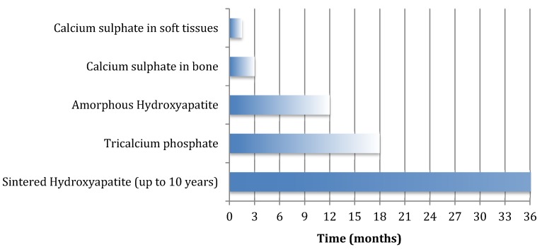
Local release of antibiotic has advantages in the treatment of chronic osteomyelitis and infected fractures. The adequacy of surgical debridement is still key to successful clearance of infection but local antibiotic carriers seem to afford greater success rates by targeting the residual organisms present after debridement and delivering much higher local antibiotic concentrations compared with systemic antibiotics alone. Biodegradable ceramic carriers can be used to fill osseous defects, which reduces the dead space and provides the potential for subsequent repair of the osseous defect as they dissolve away. A dissolving ceramic antibiotic carrier also raises the possibility of single stage surgery with definitive closure and avoids the need for subsequent surgery for spacer removal. In this article we provide an overview of the properties of various biodegradable ceramics, including calcium sulphate, the calcium orthophosphate ceramics, calcium phosphate cement and polyphasic carriers. We summarise the antibiotic elution properties as investigated in previous animal studies as well as the clinical outcomes from clinical research investigating their use in the surgical management of chronic osteomyelitis. Calcium sulphate pellets have been shown to be effective in treating local infection, although newer polyphasic carriers may support greater osseous repair and reduce the risk of further fracture or the need for secondary reconstructive surgery. The use of ceramic biocomposites to deliver antibiotics together with BMPs, bisphosphonates, growth factors or living cells is under investigation and merits further study. We propose a treatment protocol, based on the Cierny-Mader classification, to help guide the appropriate selection of a suitable ceramic antibiotic carrier in the surgical treatment of chronic osteomyelitis.

Keywords: Osteomyelitis; antibiotic carrier; biodegradable; calcium phosphate.; calcium sulphate; ceramic.


Conflict of interest statement

Competing Interests: The authors have declared that no competing interest exists.
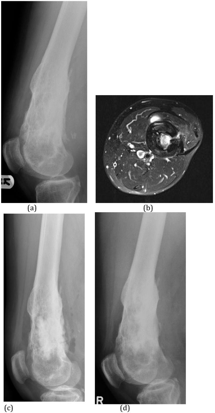
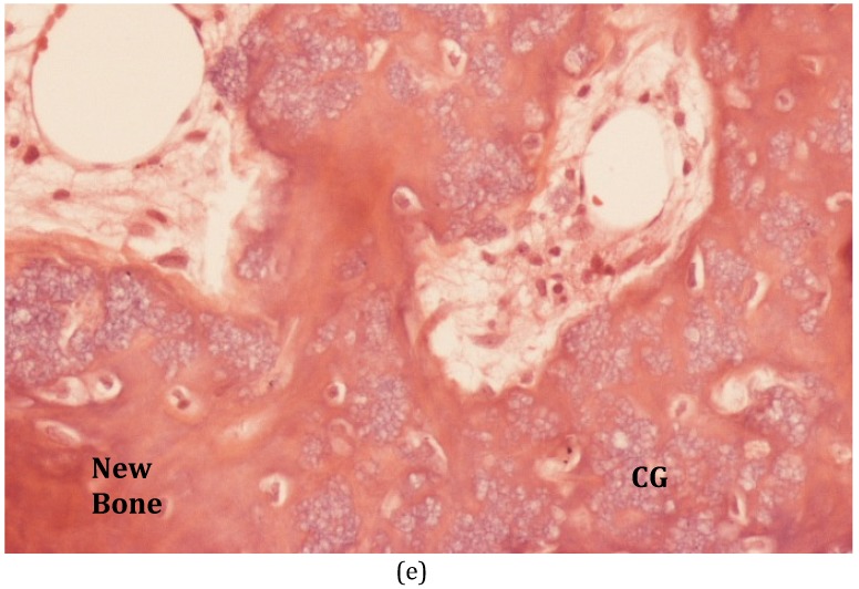
Figures

Figure 1
Figure 1
Duration of remodelling or dissolution of various ceramic biocomposites.
Figure 2
Figure 2
(A) This proximal tibial plateau fracture was treated with open reduction and internal fixation. Elevation of the depressed plateau created a bone void in the lateral condyle which was filled with a composite of α-tricalcium phosphate, calcium carbonate and monocalcium phosphate (Norian™) (white arrow). (B) Radiograph taken 4 years later shows no remodelling of this very stable ceramic. (C) The patient presented again at 6.5 years after implantation of the ceramic with an abscess over the lateral tibia (white arrows) and septic arthritis of the knee. The ceramic material remains unchanged and is now involved in the infection. It was difficult to remove at surgery.
Figure 3
Figure 3
(A) This infected ankle fusion was treated by radical excision, Ilizarov stabilisation and filling of the cortico-medullary defect with Calcium Sulphate pellets containing Tobramycin. (B) At one year after surgery, the patient is infection-free and the biocomposite has dissolved. There is almost no new bone formation within the resection defect.
Figure 4
Figure 4
(A) This diabetic patient presented with a haematogenous infection of the medullary canal of the upper femur (Cierny & Mader Type I). The MRI shows the extensive medullary oedema, intramedullary abscess and cortical involucrum. (B) The infection was excised by medullary reaming and the dead space filled with Calcium Sulphate pellets with gentamicin. (C) At 4 months after surgery, the pellets have dissolved.
Figure 5
Figure 5
(A) This patient suffered C-M Type III osteomyelitis of the distal femur after an open fracture. (B) The MRI shows the extensive medullary sequestration, the lateral cortical opening (cloaca) and anterior abscess. (C) At operation, the infected bone was excised and the cavitary defect filled with 30mls of calcium sulphate/hydroxyapatite biocomposite with gentamicin (Cerament™G). (D) Six months after implantation, the biocomposite has undergone major remodeling. (E) Bone biopsy (Haematoxylin and Eosin stained microscopy) shows widespread new bone formation within and on the surface of the material (CG = Cerament ™G).
Figure 5
Figure 5
(A) This patient suffered C-M Type III osteomyelitis of the distal femur after an open fracture. (B) The MRI shows the extensive medullary sequestration, the lateral cortical opening (cloaca) and anterior abscess. (C) At operation, the infected bone was excised and the cavitary defect filled with 30mls of calcium sulphate/hydroxyapatite biocomposite with gentamicin (Cerament™G). (D) Six months after implantation, the biocomposite has undergone major remodeling. (E) Bone biopsy (Haematoxylin and Eosin stained microscopy) shows widespread new bone formation within and on the surface of the material (CG = Cerament ™G).
Figure 6
Figure 6
(A) This infected non-union of the proximal humerus occurred after attempted minimal internal fixation with Kirschner wires. The non-union has been excised and internally fixed in a single procedure. (B) The small bone defects around the non-union, the empty holes in the plate and the central medullary space have been filled with calcium sulphate/hydroxyapatite biocomposite with gentamicin (Cerament™G).

Similar articles

Cited by

References

    1. Trampuz A, Zimmerli DW. Antimicrobial agents in orthopaedic surgery. Drugs. 2006;66(8):1089–106. - PubMed
    1. Brady RA, Leid JG, Calhoun JH, Costerton JW, Shirtliff ME. Osteomyelitis and the role of biofilms in chronic infection. Federation of European Microbiological Societies Immunology & Medical Microbiology. 2008;52(1):13–22. - PubMed
    1. Cierny G, Mader JT, Penninck JJ. The classic: A clinical staging system for adult osteomyelitis. Clinical Orthopaedics & Related Research. 2003;414:7–24. - PubMed
    1. McNally M, Nagarajah K. Osteomyelitis. Orthopaedics and Trauma. 2010;24(6):416–29.
    1. Mayberry-Carson KJ, Tober-Meyer B, Smith JK, Lambe DW, Costerton JW. Bacterial adherence and glycocalyx formation in osteomyelitis experimentally induced with staphylococcus aureus. Infection and Immunity. 1984;43(3):825–33. - PMC - PubMed