Review
. 2017 Jan 1;2(1):52-62.
doi: 10.7150/jbji.17466. eCollection 2017.

Allograft Bone as Antibiotic Carrier

Affiliations
Review

Allograft Bone as Antibiotic Carrier

Heinz Winkler et al. J Bone Jt Infect. .

Abstract

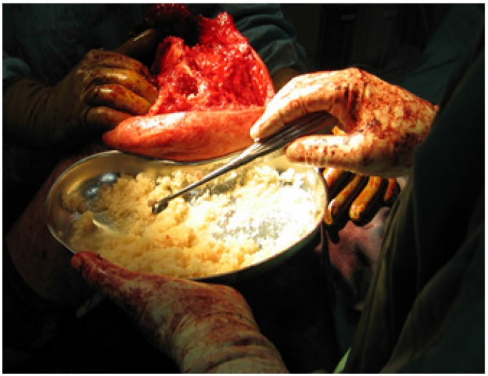
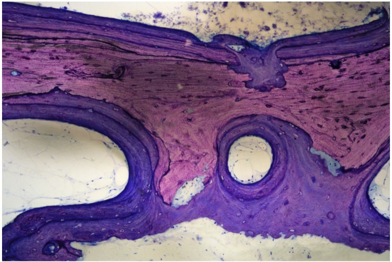
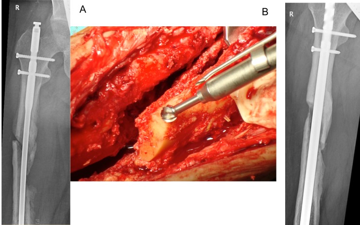
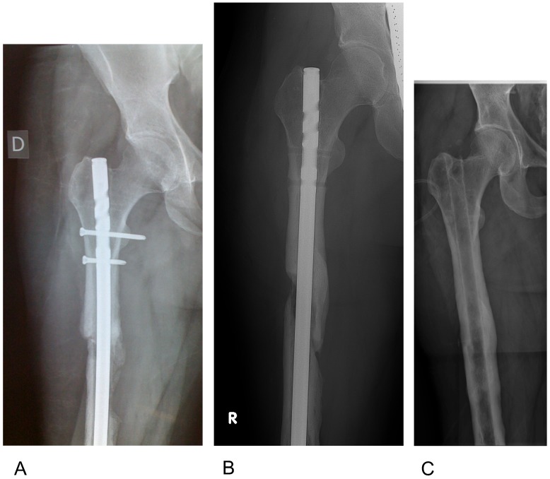
The treatment of chronic bone and joint infections is characterized by obstinate persistency of the causing microorganisms and resulting long term disability to patients, associated with remarkable costs for the health care system. Difficulties derive from biofilm formed on dead bone and eventual implants, with resistance against immunological defences and antimicrobial substances. Biofilm embedded bacteria require up to 1000 times the antibiotic concentration of planktonic bacteria for elimination. Systemic antibiotic treatment alone cannot provide the concentrations required and surgical intervention is always prerequisite for potentially providing a cure. A second issue is that osseous defects are almost always present after surgical debridement, and it is difficult to address their reconstruction. One option is to use bone grafts, either from the patient´s own body or from foreign donors (allografts). Grafts are usually unvascularized and are prone to colonization with bacteria. Loading of allografts with antibiotics may not only protect grafts from bacterial adhesion but, using appropriate processing methods, may also provide high local antibiotic concentrations that may eliminate remaining sessile pathogens. For efficient action as antibiotic carriers, the release of antibiotics should be above the minimum biofilm eradication concentration (MBEC) for a prolonged period of time. Cleaning the bone from bone marrow opens a large reservoir for storage of antimicrobial substances that, after implantation, may be released to the surrounding in a sustained mode, possibly eliminating remaining biofilm remnants. Removal of bone marrow, leaving a pure matrix, provides increased safety and improved revascularization of the graft. Local provision of antibiotic concentrations above the MBEC may enable simultaneous internal fixation with osteosynthetic material and single stage exchange of infected endoprostheses, resulting in shorter hospital stays with reduced pain and faster rehabilitation of patients.

Keywords: allograft; antibiotic carrier; biofilm; bone; infection; one stage treatment; quality of life.; reconstruction.


Conflict of interest statement

Competing Interests: The authors have declared that no competing interest exists.

Figures

Figure 1
Figure 1
A (upper figure): Unprocessed bone is filled with marrow that blocks the access to deeper layers of the available matrix surface. From: https://en.wikipedia.org/wiki/File:Spongy_bone_-_trabecules.jpg#filelinks Courtesy: Department of Histology, Jagiellonian University Medical College. B (lower figure): Bone marrow is containing immunogeneic cells and fat, eliciting undesired inflammatory reactions. Eventual pathogens may be present exclusively in the marrow, and not inside the bony matrix. From: https://commons.wikimedia.org/wiki/File%3AGray72-en.svg.
Figure 1
Figure 1
A (upper figure): Unprocessed bone is filled with marrow that blocks the access to deeper layers of the available matrix surface. From: https://en.wikipedia.org/wiki/File:Spongy_bone_-_trabecules.jpg#filelinks Courtesy: Department of Histology, Jagiellonian University Medical College. B (lower figure): Bone marrow is containing immunogeneic cells and fat, eliciting undesired inflammatory reactions. Eventual pathogens may be present exclusively in the marrow, and not inside the bony matrix. From: https://commons.wikimedia.org/wiki/File%3AGray72-en.svg.
Figure 2
Figure 2
Highly purified bone offers the advantages of 1. Increased safety, 2. Accelerated incorporation, and 3. Enormous reservoir / surface available for storage and adhesion of antimicrobial substances. Stored antibiotics are able to diffuse through the open canaliculi, providing controlled release over weeks after implantation.
Figure 3
Figure 3
Loading of carriers with antibiotics. Purified bone may store 10x the amount of vancomycin compared with cement. Almost the whole amount is available, leading to markedly elevated local concentrations and a prolonged biofilm-active release.
Figure 4
Figure 4
Antibiotic impregnated cancellous bone (Osteomyin™). Granules with a median size of 5mm in the re-hydrated condition provide a mouldable mass suitable for impaction grafting.
Figure 5
Figure 5
Biopsy of grafted OsteomycinTM, 6 months after removal of an infected dental implant and simultaneous implantation into the maxilla. “Creeping substitution” with osteoclasts resorbing the graft and osteoblasts depositing new bone simultaneously. There are no signs of inflammation.
Figure 6
Figure 6
A 24 year old male who was in a car accident with femur fracture treated with intramedullary nailing. He sustained a postoperative infection, with 3 revisions and exchange of the femoral nail. The patient presented with fever, persistent fistulation, and positive cultures (methicillin sensitive S.aureus and methicillin resistant S.epidermidis). A) Preop. X-Ray. The presence of a loose nail with unstable pseudarthrosis and major osseous defect at the non-union and around the distal locking screws (Cierny-Mader stage 4). The septic non-union was exposed and debrided. A high speed burr with stepless rotation up to 60.000rpm and integrated irrigation/cooling system provides abrasion of scleroses down to slightly bleeding bone (“Paprika-sign”), using drills of variable sizes. B) Postoperative. An exchange of the intramedullary nail was performed with rigid fixation by locking screws proximally and distally, with defects filled with antibiotic-bone-compound ABC.
Figure 7
Figure 7
Radiographical follow up of the case in Fig 6. A) 6 weeks postop: Fully weight bearing, no sign of infection. B) 1 yr postop. Dynamization was performed by removing the proximal interlocking screws; the patient is fully weight bearing with no signs of infection. C) 7yrs postop: Hardware removal. Complete union, defects restored. The patient returned to sports with no signs of infection.
Figure 8
Figure 8
A 66 year old male who sustained a femoral neck fracture treated with uncemented THR. Postoperatively he complained of unspecific pain with only slightly elevated infection markers. A) 3 years later loosening of the acetabular component was diagnosed with marked osseous defect periacetabular and signs of osteolysis around the proximal part of the stem. B) One stage exchange with uncemented components. The defects were filled with antibiotic impregnated bone Osteomycin V TM. Sonification of explanted material revealed growth of 2 strains of S.epidermidis (MSSE) and Propionibact. sp. Hospital stay was one week, with Cefuroxim intravenously, followed by 6 weeks Amoxicillin/Clavulanic acid + Rifampicin orally. C) 6 months postop: The patient is painfree with no sign of infection and unlimited mobility. There is partial remodelling of the allograft material with no sign of loosening of implants.

References

    1. Costerton JW. Biofilm theory can guide the treatment of device-related orthopaedic infections. Clin Orthop Relat Res. 2005:7–11. - PubMed
    1. Lichstein P, Gehrke T, Lombardi A. et al. One-stage vs two-stage exchange. J Arthroplasty. 2014;29:108–11. - PubMed
    1. Masquelet AC, Fitoussi F, Begue T, Muller GP. [Reconstruction of the long bones by the induced membrane and spongy autograft] Ann Chir Plast Esthet. 2000;45:346–53. - PubMed
    1. Christou C, Oliver RA, Yu Y, Walsh WR. The Masquelet technique for membrane induction and the healing of ovine critical sized segmental defects. PLoS One. 2014;9:e114122. - PMC - PubMed
    1. Karger C, Kishi T, Schneider L, Fitoussi F, Masquelet AC. Treatment of posttraumatic bone defects by the induced membrane technique. Orthop Traumatol Surg Res. 2012;98:97–102. - PubMed