Review
. 2017 Apr 21:15:215-227.
doi: 10.1016/j.nicl.2017.04.011. eCollection 2017.

Neuroimaging in pre-motor Parkinson's disease

Affiliations
Review

Neuroimaging in pre-motor Parkinson's disease

Thomas R Barber et al. Neuroimage Clin. .

Abstract

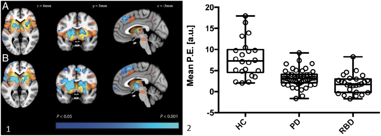
The process of neurodegeneration in Parkinson's disease begins long before the onset of clinical motor symptoms, resulting in substantial cell loss by the time a diagnosis can be made. The period between the onset of neurodegeneration and the development of motoric disease would be the ideal time to intervene with disease modifying therapies. This pre-motor phase can last many years, but the lack of a specific clinical phenotype means that objective biomarkers are needed to reliably detect prodromal disease. In recent years, recognition that patients with REM sleep behaviour disorder (RBD) are at particularly high risk of future parkinsonism has enabled the development of large prodromal cohorts in which to investigate novel biomarkers, and neuroimaging has generated some of the most promising results to date. Here we review investigations undertaken in RBD and other pre-clinical cohorts, including modalities that are well established in clinical Parkinson's as well as novel imaging methods. Techniques such as high resolution MRI of the substantia nigra and functional imaging of Parkinsonian brain networks have great potential to facilitate early diagnosis. Further longitudinal studies will establish their true value in quantifying prodromal neurodegeneration and predicting future Parkinson's.


Figures

Fig. 1
Fig. 1
Serial SPECT images from two patients (A–C and D–F) with RBD, showing labelling of the basal ganglia with 123I-FP-CIT. Images are at baseline (A and D), 1.5 years (B and E) and 3 years (C and F). A greater signal decline over time is seen in the patient who developed Parkinson's (D–E) than in the patient who did not (A–C).
Fig. 2
Fig. 2
Transcranial ultrasound (TCS) of the midbrain. A: axial T1-weighted MRI at the level of the midbrain demonstrating the approximate area imaged by TCS in B and C. B: TCS of the midbrain (outlined) in an individual with normal appearances of the substantia nigra (SN). Arrowheads indicate signal from the surrounding basal cisterns. R = raphe. C: TCS from an individual with hyperechogenicity of the substantia nigra (SN, encircled within the midbrain outline). White arrows indicate the position of the red nuclei.
Fig. 3
Fig. 3
Clinical high resolution 3D–T2*/SWI MRI of the midbrain showing the normal appearance of the substantia nigra (right image) with high intensity signal in the dorsolateral region, and absence of this feature (left image) in a patient with Parkinson's.
Fig. 4
Fig. 4
A: representation of the Parkinson's Disease Related spatial covariance pattern (PDRP) derived from FDG-PET imaging, with increased metabolic activity denoted in red-yellow and decreased metabolic activity in blue-green. B: baseline PDRP expression levels in 20 individuals with RBD, divided into those who remained disease free (CON(−)) and those went on to develop MSA or PD/DLB. C: A combination of PDRP expression and age was able to separate the three groups into distinct clusters. (For interpretation of the references to color in this figure legend, the reader is referred to the online version of this chapter.)
Fig. 5
Fig. 5
Resting state MRI in individuals with RBD, PD and healthy controls. Panel 1 (left): resting state functional connectivity in PD (A) and RBD (B). The basal ganglia network template derived from a separate group of control participants is shown in red-yellow. Areas where functional connectivity was significantly reduced compared to controls is shown in blue. Panel 2 (right): extracted mean parameter estimates from the clusters with significantly different connectivity in both the PD versus control and RBD versus control comparisons. Box plots represent the group mean and quartiles with whiskers denoting minimum and maximum values. (For interpretation of the references to color in this figure legend, the reader is referred to the online version of this chapter.)

References

    1. https://www.cureparkinsons.org.uk/trials-repurposed-drugs
    1. Adams J.R., van Netten H., Schulzer M. PET in LRRK2 mutations: comparison to sporadic Parkinson's disease and evidence for presymptomatic compensation. Brain. 2005;128:2777–2785. - PubMed
    1. Albin R.L., Koeppe R.A., Chervin R.D., Consens F.B., Wernette K., Frey K.A. Decreased striatal dopaminergic innervation in REM sleep behavior disorder. Neurology. 2000;55(9):1410–1412. - PubMed
    1. Arnaldi D., de Carli F., Picco A., Ferrara M., Accardo J., Bossert I. Nigro-caudate dopaminergic deafferentation: a marker of REM sleep behavior disorder? Neurobiol. Aging. 2015;36(12):3300–3305. - PubMed
    1. Barkhof F., Haller S., Rombouts S.A. Resting-state functional MR imaging: a new window to the brain. Radiology. 2014 Jul;272(1):29–49. - PubMed

Publication types

MeSH terms