

Editorial
doi: 10.21037/tlcr.2017.04.09.
Cons: the confusing mucinous adenocarcinoma classification
Affiliations
- PMID: 28529906
- PMCID: PMC5420532
- DOI: 10.21037/tlcr.2017.04.09
Item in Clipboard
Editorial
Cons: the confusing mucinous adenocarcinoma classification
Transl Lung Cancer Res.
2017 Apr.
No abstract available
Conflict of interest statement
Conflicts of Interest: The author has no conflicts of interest to declare.
Figures
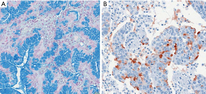
Examples of invasive mucinous adenocarcinomas. (A) Invasive mucinous acinar adenocarcinoma, goblet cell type; it is evident from the figure that most tumor cells have produced mucins and stored this in the cytoplasmic vacuoles, however, some tumor cells are releasing mucin into the acinar lumen, whereas few others have started new synthesis; (B) staining by antibodies for MUC5AC shows different states of synthesis as well as release of the material into the lumen. Bar 20 µm.
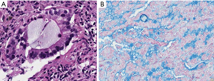
Invasive mucinous adenocarcinoma. In both (A) and (B) the vast majority of tumor cells produce and secrete mucins, however, in both also the different stages of mucin synthesis and secretion can be seen. Cells having already released mucin are smaller, even flat, whereas tumor cells storing mucin are large cuboidal. H&E and Alcian blue, bars 10 and 20 µm.
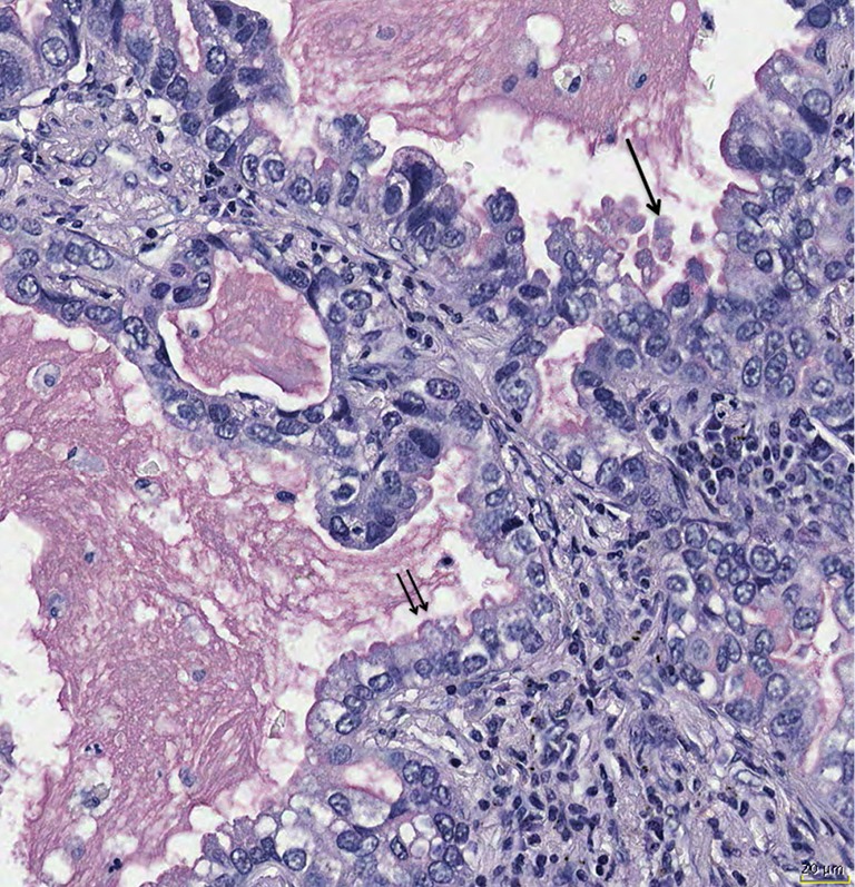
Mucin secretion can show variations; holocrine secretion is characterized by release of mucin together with vesicles from the cytoplasm (arrow), whereas apocrine secretion is secretion into the lumen without cytoplasmic material (double arrow). Mucicarmine stain, bar 20 µm.
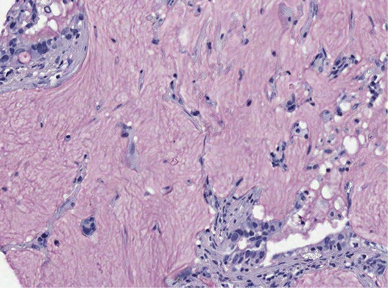
Colloid adenocarcinoma, the tumor cells secrete mucins from every side, therefore they easily detach from the tumor complex and float within the mucin. Even in this type of carcinoma there is no synchronized mucin synthesis and release. Note also few signet ring cells in the upper left corner. Mucicarmine stain, ×200.
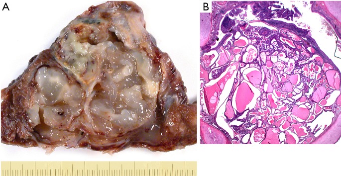
Examples of invasive mucinous cystadenocarcinoma (A) and in situ cystadenocarcinoma (B). (A) Mucinous cystadenocarcinoma, resected specimen showing the thin fibrous pseudocapsule; in (B) a precursor lesion, mucinous cystadenoma, borderline variant is shown. This in essence is an in situ mucinous cystadenocarcinoma. H&E ×25.
References
-
- Travis WD, Brambilla E, Burke AP, et al. WHO Classification of Tumours of the Lung, Pleura, Thymus and Heart. Fourth edition ed. WHO Classification of Tumours. Geneva: IARC, WHO Press, 2015. - PubMed
-
- Travis WD, Brambilla E, Noguchi M, et al. International association for the study of lung cancer/american thoracic society/european respiratory society international multidisciplinary classification of lung adenocarcinoma. J Thorac Oncol 2011;6:244-85. 10.1097/JTO.0b013e318206a221 - DOI - PMC - PubMed
-
- Kadota K, Yeh YC, Sima CS, et al. The cribriform pattern identifies a subset of acinar predominant tumors with poor prognosis in patients with stage I lung adenocarcinoma: a conceptual proposal to classify cribriform predominant tumors as a distinct histologic subtype. Mod Pathol 2014;27:690-700. 10.1038/modpathol.2013.188 - DOI - PMC - PubMed
-
- Travis W, Colby TV, Corrin B, et al. Histological Typing of Lung and Pleura Tumours. 3rd ed. WHO, International Histological Classification of Tumours. Berlin, Heidelberg, New York: Spinger; 1999.
Publication types
LinkOut - more resources
Full Text Sources