Review
. 2017 May 5:4:24.
doi: 10.3389/fcvm.2017.00024. eCollection 2017.

Coronary Artery Aneurysms: A Review of the Epidemiology, Pathophysiology, Diagnosis, and Treatment

Affiliations
Review

Coronary Artery Aneurysms: A Review of the Epidemiology, Pathophysiology, Diagnosis, and Treatment

Sara Abou Sherif et al. Front Cardiovasc Med. .

Abstract

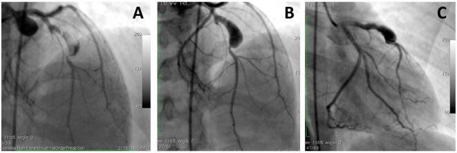
Coronary artery aneurysms (CAAs) are uncommon and describe a localized dilatation of a coronary artery segment more than 1.5-fold compared with adjacent normal segments. The incidence of CAAs varies from 0.3 to 5.3%. Ever since the dawn of the interventional era, CAAs have been increasingly diagnosed on coronary angiography. Causative factors include atherosclerosis, Takayasu arteritis, congenital disorders, Kawasaki disease (KD), and percutaneous coronary intervention. The natural history of CAAs remains unclear; however, several recent studies have postulated the underlying molecular mechanisms of CAAs, and genome-wide association studies have revealed several genetic predispositions to CAA. Controversies persist regarding the management of CAAs, and emerging findings support the importance of an early diagnosis in patients predisposed to CAAs, such as in children with KD. This review aims to summarize the present knowledge of CAAs and collate the recent advances regarding the epidemiology, etiology, pathophysiology, diagnosis, and treatment of this disease.

Keywords: atherosclerosis; coronary aneurysm; coronary artery aneursym; coronary artery disease; coronary artery ectasia; coronary stent.


Figures

Figure 1
Figure 1
Acute coronary syndrome and coronary artery aneurysms. A patient presented with acute coronary aneurysm. (A,B) Coronary angiography revealed an aneurysmal dilatation of the ostia of the left anterior descending (LAD) artery and left circumflex artery, with a thrombus image (arrow heads) in the LAD artery along with a significant stenosis in the proximal part. Control angiogram failed to show any reduction in the thrombus after 48 h of tirofiban infusion, and the patient was referred to the surgery. (C) Aneurysmal dilatation can also be seen in computed tomography images.
Figure 2
Figure 2
A large aneurysm in left anterior descending artery in a 17-year-old patient with neurofibromatosis causing acute coronary syndrome. Coronary angiograms with (A) and without (B,C) thrombus image in the aneurysm after anticoagulation. However, this patient was referred to surgery to prevent future complications [with permission from the report by Evrengul et al. (92)].
Figure 3
Figure 3
A patient with coronary aneurysm due to Kawasaki disease. (A) Computed tomography angiography showed a large aneurysm (maximum diameter 9 mm, extending for more than 35 mm) of the proximal left anterior descending (LAD) artery with calcific degeneration, large filling defect with thrombus and severe lumen narrowing. (B) Coronary angiography confirmed a 70% stenosis of the LAD artery with an aneurysm immediately distal to the ostium (maximum diameter 9.2 mm × 7.8 mm). (C) Final angiography demonstrating exclusion of the aneurysm using a covered stent [with permission from the report by Di Mario et al. (133)].
Figure 4
Figure 4
A 70-year-old male presented with angina on exertion. Three years prior to this presentation, they had an abdominal aortic aneurysm repair.(A,B) Coronary angiogram revealed coronary artery disease and an aneurysm in the left anterior descending artery. The patient was referred to surgery, because of the risk of occlusion of the significant side branches, if a stent was to be used. (C,D) Coronary aneurysm was surgically excluded with a linear repair (images courtesy of Yalin Tolga Yaylali and Bilgin Emrecan).

References

    1. Markis JE, Joffe CD, Cohn PF, Feen DJ, Herman MV, Gorlin R. Clinical significance of coronary arterial ectasia. Am J Cardiol (1976) 37(2):217–22. 10.1016/0002-9149(76)90315-5 - DOI - PubMed
    1. Topaz O, Rutherford MS, Mackey-Bojack S, Prinz AW, Katta S, Salter D, et al. Giant aneurysms of coronary arteries and saphenous vein grafts: angiographic findings and histopathological correlates. Cardiovasc Pathol (2005) 14:298–302. 10.1016/j.carpath.2005.08.007 - DOI - PubMed
    1. Hartnell GG, Parnell BM, Pridie RB. Coronary artery ectasia: its prevalence and clinical significance in 4993 patients. Br Heart J (1985) 54:392–5. 10.1136/hrt.54.4.392 - DOI - PMC - PubMed
    1. Packard M, Wechsler HF. Aneurysms of coronary arteries. Arch Intern Med (1929) 43:1–14. 10.1001/archinte.1929.00130240004001 - DOI
    1. Robertson T, Fisher L. Prognostic significance of coronary artery aneurysm and ectasia in the Coronary Artery Surgery Study (CASS) registry. Prog Clin Biol Res (1987) 250:325–39. - PubMed

LinkOut - more resources