

Effect of intraoperative positioning on the diameter of the vertebral canal in cats during perineal urethrostomy (cadaveric study)
- PMID: 28530134
- PMCID: PMC11129205
- DOI: 10.1177/1098612X17709645
Effect of intraoperative positioning on the diameter of the vertebral canal in cats during perineal urethrostomy (cadaveric study)
Abstract
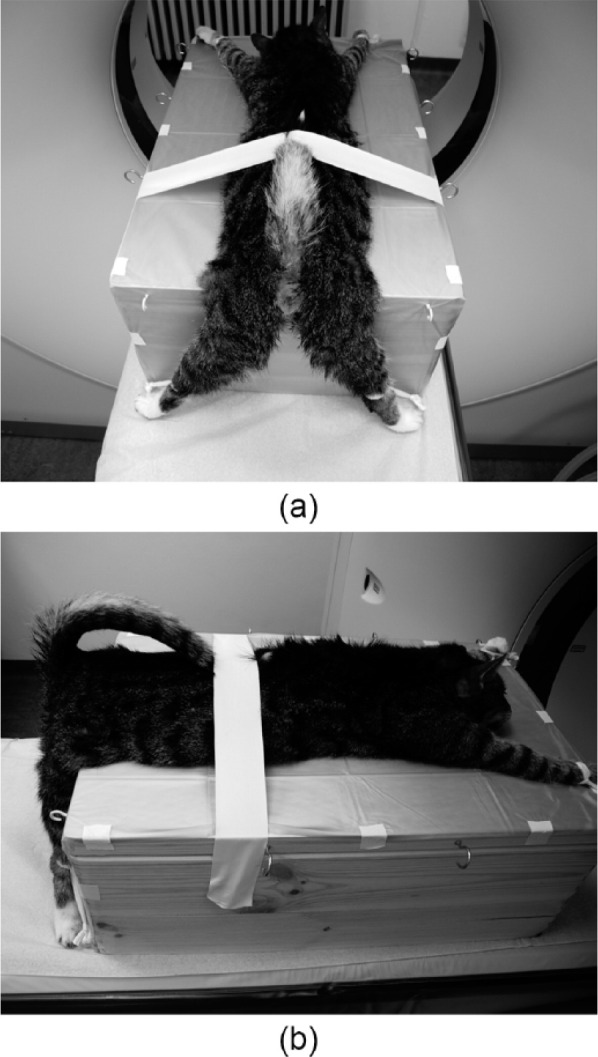
Objectives The objective of this study was to quantify the changes in the diameter of the vertebral canal in the lumbosacral and sacrococcygeal column (L6-Co2) in cats in dorsal and ventral recumbency, simulating real body positioning during a perineal urethrostomy. Methods Twenty-one male feline cadavers were enrolled in the study. All feline cadavers were evaluated by CT. Examinations were performed with the cadaver in a neutral position and dorsal and ventral recumbency. Sagittal vertebral canal diameters (VCDs) were obtained by measuring the distance between the ventral and dorsal aspects of the vertebral canal in the middle of the intervertebral space. Results A comparison of the VCDs between L6 and L7, L7 and S1, S3 and Co1 and Co1 and Co2 in neutral position vs dorsal recumbency revealed a reduction of 0.27 mm (14.6%; P <0.001) between S3 and Co1 and 0.26 mm (18.1%; P <0.001) between Co1 and Co2. No differences were seen when comparing L6-L7 and L7-S1. The VCDs were decreased in all segments when comparing neutral with ventral recumbency. This study revealed a reduction of 0.13 mm between L6 and L7 (3.3%; P = 0.003), 0.14 mm between L7 and S1 (4.1%; P = 0.003), 0.61 mm between S3 and Co1 (32.5%; P <0.001) and 0.63 mm between Co1 and Co2 (44.1%; P <0.001). Comparison of the VCD between dorsal and ventral recumbency in L6-L7, L7-S1, S3-Co1 and Co1-Co2 revealed a decrease in the VCDs in ventral recumbency of 0.13 mm (3.3%; P <0.001), 0.12 mm (3.6%; P <0.001), 0.34 mm (21.0%; P <0.001) and 0.37 mm (31.7%; P <0.001), respectively. Conclusions and relevance The results provide evidence that, from an anatomical point of view, perineal urethrostomy performed in dorsal recumbency is superior to ventral recumbency, but further clinical studies to verify these findings are necessary.
Conflict of interest statement
The authors declared no potential conflicts of interest with respect to the research, authorship, and/or publication of this article.
Figures


References
-
- Blake JA. Perineal urethrostomy in cats. J Am Vet Med Assoc 1968; 152: 1499–1506. - PubMed
-
- Carbone MG. A modified technique for perineal urethrostomy in the male cat. J Am Vet Med Assoc 1967; 151: 301–305. - PubMed
-
- Johnston DE. Feline urethrostomy – a critique and new method. J Small Anim Pract 1974; 15: 421–435. - PubMed
-
- Richards DA, Hinko PJ, Morse EM, Jr. Feline perineal urethrostomy: a new technique for an old problem. J Am Anim Hosp Assoc 1972; 8: 66–73.
MeSH terms
LinkOut - more resources
Full Text Sources
Other Literature Sources
Miscellaneous
