. 2017 May 22:23:2436-2444.
doi: 10.12659/msm.901926.

Visualization of Penile Suspensory Ligamentous System Based on Visible Human Data Sets

Affiliations

Visualization of Penile Suspensory Ligamentous System Based on Visible Human Data Sets

Xianzhuo Chen et al. Med Sci Monit. .

Abstract

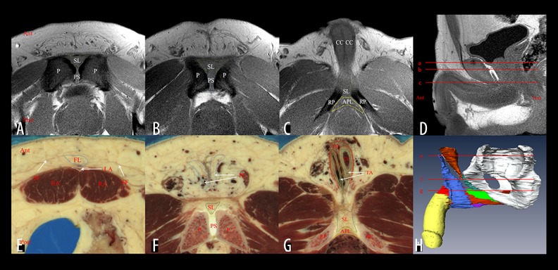
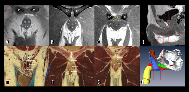
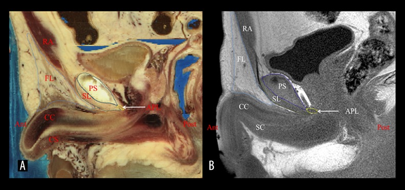
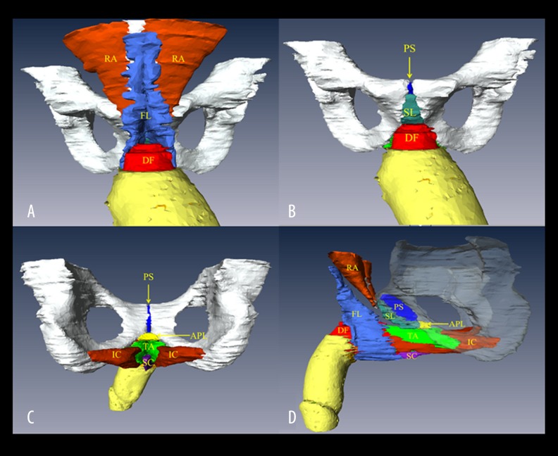
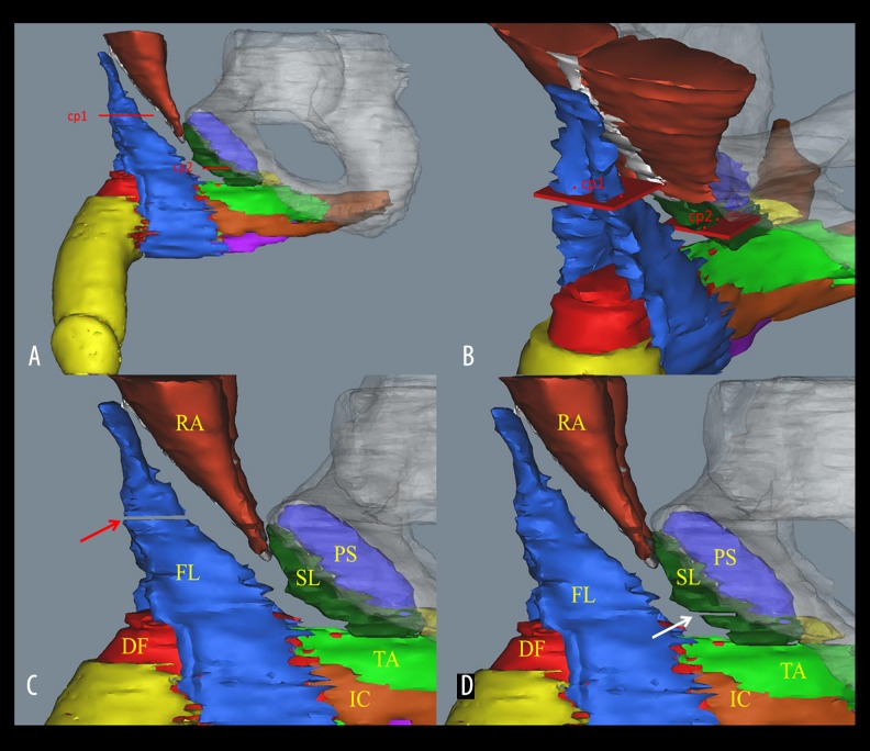
BACKGROUND The aim of this study was to use a three-dimensional (3D) visualization technology to illustrate and describe the anatomical features of the penile suspensory ligamentous system based on the Visible Human data sets and to explore the suspensory mechanism of the penis for the further improvement of the penis-lengthening surgery. MATERIAL AND METHODS Cross-sectional images retrieved from the first Chinese Visible Human (CVH-1), third Chinese Visible Human (CVH-3), and Visible Human Male (VHM) data sets were used to segment the suspensory ligamentous system and its adjacent structures. The magnetic resonance imaging (MRI) images of this system were studied and compared with those from the Visible Human data sets. The 3D models reconstructed from the Visible Human data sets were used to provide morphological features of the penile suspensory ligamentous system and its related structures. RESULTS The fundiform ligament was a superficial, loose, fibro-fatty tissue which originated from Scarpa's fascia superiorly and continued to the scrotal septum inferiorly. The suspensory ligament and arcuate pubic ligament were dense fibrous connective tissues which started from the pubic symphysis and terminated by attaching to the tunica albuginea of the corpora cavernosa. Furthermore, the arcuate pubic ligament attached to the inferior rami of the pubis laterally. CONCLUSIONS The 3D model based on Visible Human data sets can be used to clarify the anatomical features of the suspensory ligamentous system, thereby contributing to the improvement of penis-lengthening surgery.


Conflict of interest statement

Conflict of interest

The authors have no conflict of interest to declare in regard to this work.

Figures

Figure 1
Figure 1
Cross-sectional images of the suspensory ligamentous system and its adjacent structures on the VHM and MRI images. (D) Positions of the transverse sections of panel A–C. Sections show the medium-signal-intensity suspensory ligament (A, B) and arcuate pubic ligament (C) on the MRI images. (H) Positions of the transverse sections of panel E–G. Sections show the position of the fundiform ligament (A, B), suspensory ligament (B), and arcuate pubic ligament (C) in VHM data set. SF – Scarpa’s fascia; FL – fundiform ligament; LA – linea alba; RA – rectus abdominis; SL – suspensory ligament; PS – pubic symphysis; P – pubis; DF – deep fasica of the penis; CC – corpora cavernosa; TA – tunica albuginea; APL – arcuate pubic ligament; RP – ramus of pubis.
Figure 2
Figure 2
Coronal section of the suspensory ligamentous system and its adjacent structures from the front on the VHM and MRI images. (D) Positions of the coronal sections of panel A–C. Sections show the 2 high-signal intensity bundles of the fundiform ligament (A), the medium-signal intensity suspensory ligament (B) and the medium-signal intensity arcuate pubic ligament (C) on MRI images. (H) Positions of the coronal sections of panel E–G. Sections show the 2 bundles of the fundiform ligament (E), the suspensory ligament (F), and the arcuate pubic ligament (G) in VHM data set. FL – fundiform ligament; LA – linea alba; RA – rectus abdominis; SL – suspensory ligament; PS – pubic symphysis; P – pubis; DF – deep fasica of the penis; CC – corpora cavernosa; CS – corpus spongiosum; TA – tunica albuginea; APL – arcuate pubic ligament; RP – ramus of pubis.
Figure 3
Figure 3
Mid-sagittal section of the suspensory ligament and its related structures on the VHM and MRI images. (A) Mid-sagittal section of the suspensory ligamentous system and its related structures in the VHM data set. (B) Mid-sagittal section MRI image of the suspensory ligamentous system and its related structures. FL – fundiform ligament; RA – rectus abdominis; SL – suspensory ligament; PS – pubic symphysis; CC – corpora cavernosa; CS – corpus spongiosum; TA – tunica albuginea; APL – arcuate pubic ligament.
Figure 4
Figure 4
Visualization of the suspensory ligamentous system and its related structures based on the VHM data set. (A) Anterior view of the 3D model. (B) The anterior view of the 3D model with removing of the fundiform ligament, linea alba and rectus abdominis. (C) Posterior view of the 3D model. (D) Left lateral view of the 3D model with transparence of the pelvic bone. FL – fundiform ligament; LA – linea alba; RA – rectus abdominis; SL – suspensory ligament; PS – pubic symphysis; DF – deep fasica of the penis; CC – corpora cavernosa; TA – tunica albuginea; APL – arcuate pubic ligament; IC – ischiocavernosus muscle.
Figure 5
Figure 5
3D illustration of the dissection of the penile ligaments. (A) Left lateral view of the cut plane of the penile ligaments; (B) the isometric view of the cut plane of the penile ligaments; (C) the fundiform ligament is completely dissected at the level of the superior border of the pubic symphysis (the red arrow); and (D) the suspensory ligament is partially dissected (white arrow). Depth of incision reaches the anteroinferior border of pubic arch. cp1 – the cut plane of the fundiform ligament; cp2 – the cut plane of the suspensory ligament; FL – fundiform ligament; RA – rectus abdominis; SL – suspensory ligament; PS – pubic symphysis; DF – deep fasica of the penis; TA – tunica albuginea; IC – ischiocavernosus muscle.

Similar articles

Cited by

References

    1. Li CY, Agrawal V, Minhas S, Ralph DJ. The penile suspensory ligament: Abnormalities and repair. BJU Int. 2007;99:117–20. - PubMed
    1. Spyropoulos E, Christoforidis C, Borousas D, et al. Augmentation phalloplasty surgery for penile dysmorphophobia in young adults: Considerations regarding patient selection, outcome evaluation and techniques applied. Eur Urol. 2005;48:121–28. - PubMed
    1. Li CY, Kayes O, Kell PD, et al. Penile suspensory ligament division for penile augmentation: indications and results. Eur Urol. 2006;49:729–33. - PubMed
    1. Yongsheng S, Qingping Y, Yiyang J, et al. [Clinical experience of penile elongation: A comparison of four different operative approaches]. Zhonghua Zheng Xing Wai Ke Za Zhi. 2015;31:411–13. [in Chinese] - PubMed
    1. Egydio PH, Kuehhas FE. Penile lengthening and widening without grafting according to a modified ‘sliding’ technique. BJU Int. 2015;116:965–72. - PubMed