. 2017 May 22:8:15395.
doi: 10.1038/ncomms15395.

TWEAK mediates inflammation in experimental atopic dermatitis and psoriasis

Affiliations

TWEAK mediates inflammation in experimental atopic dermatitis and psoriasis

Daniel Sidler et al. Nat Commun. .

Abstract

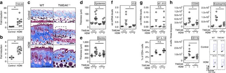
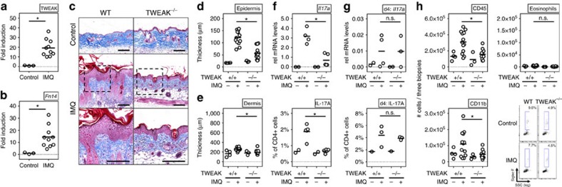
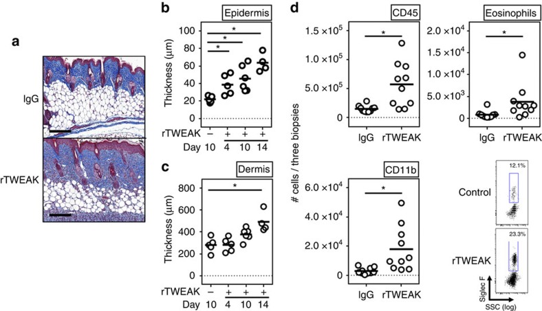
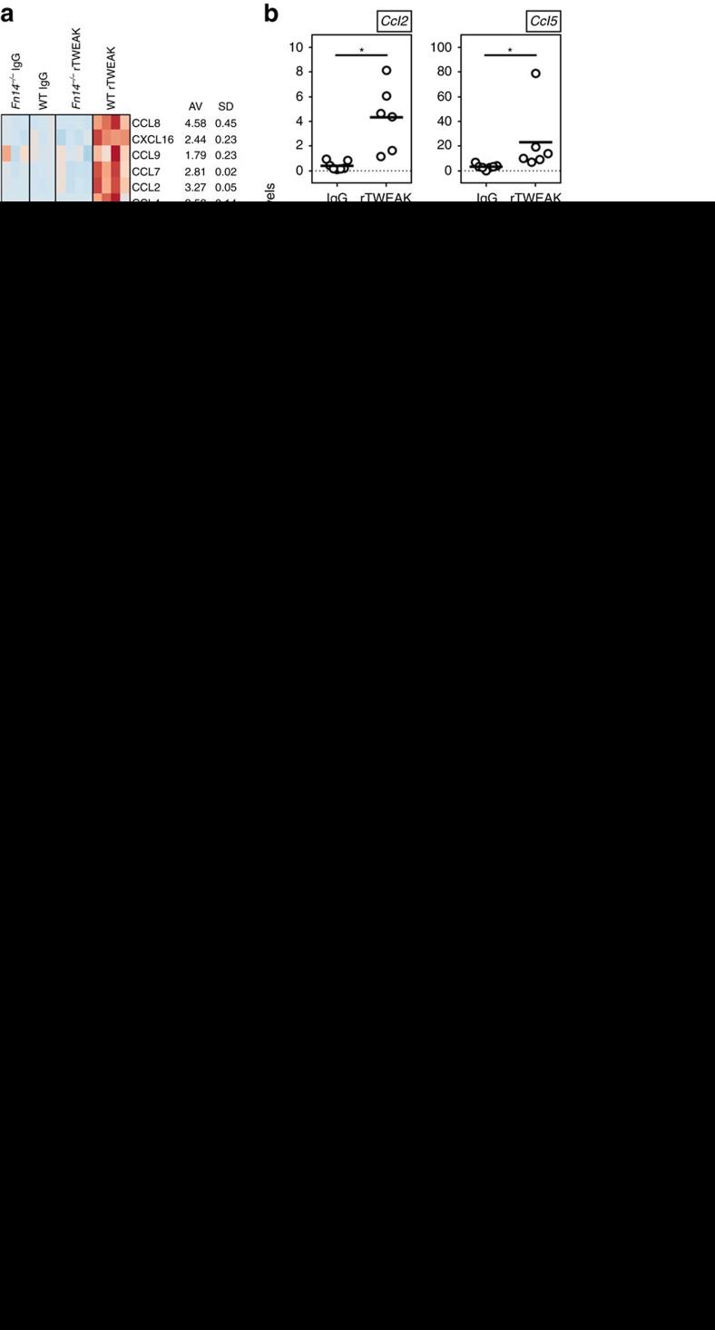
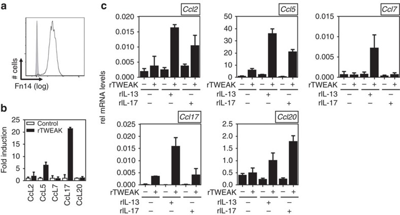
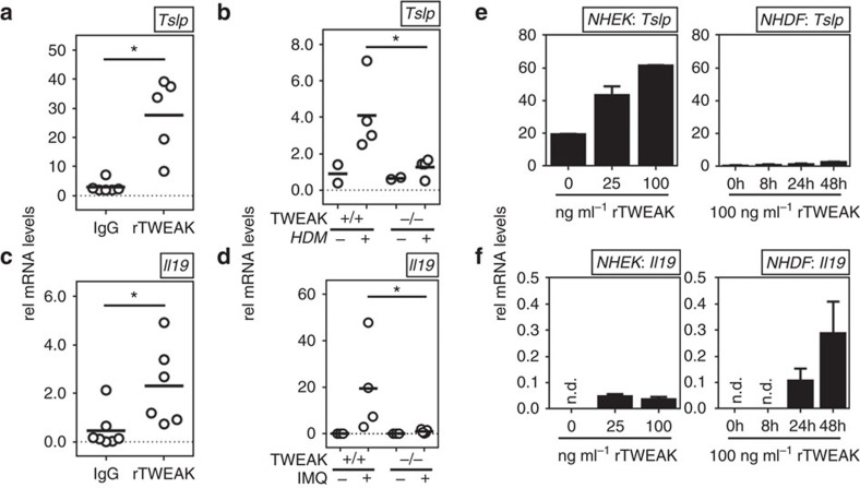
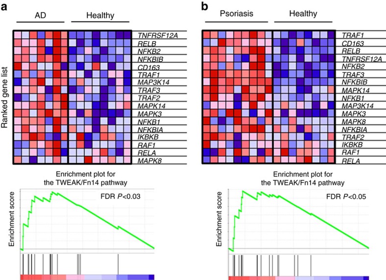
Atopic dermatitis (AD) and psoriasis are driven by alternate type 2 and type 17 immune responses, but some proteins might be critical to both diseases. Here we show that a deficiency of the TNF superfamily molecule TWEAK (TNFSF12) in mice results in defective maintenance of AD-specific T helper type 2 (Th2) and psoriasis-specific Th17 cells in the skin, and impaired expression of disease-characteristic chemokines and cytokines, such as CCL17 and TSLP in AD, and CCL20 and IL-19 in psoriasis. The TWEAK receptor, Fn14, is upregulated in keratinocytes and dermal fibroblasts, and TWEAK induces these cytokines and chemokines alone and in synergy with the signature T helper cytokines of either disease, IL-13 and IL-17. Furthermore, subcutaneous injection of recombinant TWEAK into naive mice induces cutaneous inflammation with histological and molecular signs of both diseases. TWEAK is therefore a critical contributor to skin inflammation and a possible therapeutic target in AD and psoriasis.


Conflict of interest statement

The authors declare no competing financial interests.

Figures

Figure 1
Figure 1. TWEAK controls disease activity in HDM-induced AD.
WT and TWEAK-deficient mice were treated epicutaneously with HDM on the shaved back for 23 days to induce AD-like disease. (a,b) TWEAK and Fn14 mRNA fold induction in WT mice relative to expression in skin from naive animals (Control). Combined results from three experiments. Each data point represents one individual mouse. (c) Representative Masson's trichrome staining of skin sections from WT and TWEAK−/− animals treated with HDM or PBS control. Bar represents 200 μm. (d,e) Epidermal and dermal thickness measured on trichrome-stained sections. Results combined from three independent experiments. Each data point represents one animal. (f) IL-13 mRNA in skin lesions on day 23. Combined from two experiments. (g) IL-13 mRNA in skin biopsies (top), and frequency of IL-13 positive CD4+ T cells in skin-draining lymph nodes on day 7 (bottom). Four mice per group. (h) Total numbers of CD45+ cells (left top), CD45+CD11b+ monocytes (left bottom), and SigF+CD45+CD11b+Ly6G eosinophils (right top and bottom) in skin biopsies on day 23. Combined results from two experiments, with each data point one animal. Bars represent mean values. The Mann–Whitney U-test was performed to compare the groups. Asterisks indicate P<0.05. n.s. indicates nonsignificant differences.
Figure 2
Figure 2. Reduced disease severity in IMQ-induced psoriasis in the absence of TWEAK.
WT and TWEAK-deficient mice were treated epicutaneously on the shaved back with IMQ for 8 days to induce psoriasis-like disease. (a,b) TWEAK and Fn14 mRNA fold induction in WT mice relative to expression in skin from naive animals (control). Combined results from three experiments. Each data point an individual mouse. (c) Representative Masson's trichrome staining of skin sections. Scale bar, 200 μm. (d,e) Epidermal and dermal thickness on day 8 measured on trichrome-stained sections. (f,g) IL-17 mRNA in skin lesions and frequency of IL-17 positive CD4+ T cells in skin-draining lymph nodes on day 8 (f) or day 4 (g). Each datapoint one mouse. (h) Total numbers of CD45+ cells (left top), CD45+CD11b+ monocytes (left bottom) and SigF+CD45+CD11b+Ly6G eosinophils (right) from skin biopsies obtained on day 8. Combined results from three experiments. Each data point one animal. Bars represent mean values. The Mann–Whitney U-test was performed to compare the groups. Asterisks indicate P<0.05. n.s. indicates nonsignificant differences.
Figure 3
Figure 3. rTWEAK induces skin inflammation.
(a,b) Naive WT mice were injected with 75 μg mouse rTWEAK or IgG, administered s.c. either four times during a 14 day period (a), or once with analysis on day 4, or twice weekly with analysis on day 10 (b,c). (a) Masson's trichrome staining of representative skin sections on day 14. Scale bar, 200 μm. (b,c) Epidermal and dermal thickness on days 4, 10 and 14. Results are combined from two independent experiments. (d) Absolute number of skin-infiltrating cells measured by flow cytometry on day 4, and representative flow plot of skin-infiltrating eosinophils (SigF+CD45+CD11b+Ly6G cells). Combined from two experiments. Each data point one animal. Bars represent mean values. The ANOVA test with Bonferroni Correction was performed to compare the groups. Asterisks indicate P<0.05. n.s. indicates nonsignificant differences. ANOVA, analysis of variance.
Figure 4
Figure 4. TWEAK regulates chemokine expression in the skin.
(a,b) Naive WT or Fn14-deficient mice were injected with rTWEAK or IgG, administered once s.c. After 4 days expression of mRNA for indicated chemokines was measured in skin biopsies. (a, left) RNAseq heatmap of chemokine expression from 3 to 4 animals/group. (right) Mean (AV) fold increase and SD calculated from RNAseq data of the comparisons of rTWEAK into WT versus Fn-deficient mice, rTWEAK versus IgG into WT mice, and rTWEAK versus no injection into WT mice. (b) qPCR mRNA expression of indicated chemokines relative to GAPDH. Combined results from two experiments. Each data point one mouse. (cf) naive WT mice were injected with rTWEAK or IgG (s.c.) twice weekly over 14 days as in Fig. 3a, with either a combination of blocking antibodies to CCL2, CCL5 and CCL7, or a combination of control antibodies (i.p). Combined results from two experiments. (c) Masson's trichrome staining of representative skin sections. Scale bar, 200 μm. (d,e) Quantification of epidermal and dermal thickness. (f) Total numbers of CD45+ cells and SigF+CD45+CD11b+Ly6G eosinophils from skin biopsies. (g,h) WT and TWEAK-deficient animals were treated with HDM for 23 days (g) and IMQ for 8 days (h) and mRNA for indicated chemokines assessed in skin, relative to GAPDH expression. Combined from two experiments. Each data point one mouse. Bars represent mean values. The Mann–Whitney U-test was performed to compare the groups. Asterisks indicate P<0.05. qPCR, quantitative PCR.
Figure 5
Figure 5. TWEAK synergizes with IL-13 and IL-17A to induce chemokines in human keratinocytes.
(a) Basal surface expression of Fn14 (line) on NHEK cells relative to Isotype staining (grey). (b,c) NHEK cells were stimulated for 48 h with rTWEAK, rIL-13, rIL-17 or their combination. mRNA expression of indicated chemokines was assessed relative to GAPDH. Means of triplicate cultures with standard deviations. Values were compared using Student's t-test. One out of three independent experiments.
Figure 6
Figure 6. TWEAK promotes cytokines associated with AD and psoriasis.
(a,c) Naive WT mice were injected with rTWEAK or IgG administered once s.c. After 4 days expression of mRNA for TSLP and IL-19 was measured in skin biopsies. Data relative to GAPDH. Combined results from two experiments. (b,d) WT and TWEAK-deficient animals were treated with HDM for 4 days (b) or IMQ for 8 days (d) and mRNA for TSLP and IL-19, respectively, assessed in skin, relative to GAPDH expression. One out of three experiments shown. (e,f) NHEK cells (left) or NHDF cells (right) were stimulated for 48 h or the indicated times with rTWEAK. mRNA expression of TSLP (e) and IL-19 (f) was assessed relative to GAPDH. Each data point in ad represents one mouse. Bars represent mean values. The Mann–Whitney U-test was performed to compare the groups. Bars in (e,f) indicate mean values of triplicates and s.d. Values were compared using Student's t-test. One out of three independent experiments. Asterisks indicate P<0.05.
Figure 7
Figure 7. Gene set enrichment for the TWEAK/Fn14 pathway in AD and psoriasis.
Gene set enrichment analysis of skin biopsies from healthy (n=8), AD (n=7), and psoriasis patients (n=9). GenePattern 2.0 (ref. 74) was used to process RNA array data for gene set enrichment analysis which was run with the default settings (100 iterations, weighted). A FDR P value <0.05 was considered significant. Fn14 (TNFRSF12A) and intracellular signalling and effector molecules are significantly enriched among the induced transcripts from AD (a) and psoriasis lesions (b) when compared to healthy control specimens (FDR P<0.05 in both comparisons). FDR, false discovery rate.

Similar articles

Cited by

References

    1. Bieber T. Atopic dermatitis. N. Engl. J. Med. 358, 1483–1494 (2008). - PubMed
    1. Nestle F. O., Kaplan D. H. & Barker J. Psoriasis. N. Engl. J. Med. 361, 496–509 (2009). - PubMed
    1. Ring J., Mohrenschlager M. & Weidinger S. Molecular genetics of atopic eczema. Chem. Immunol. Allergy 96, 24–29 (2012). - PubMed
    1. Bieber T. & Novak N. Pathogenesis of atopic dermatitis: new developments. Curr. Allergy Asthma Rep. 9, 291–294 (2009). - PubMed
    1. Harden J. L., Krueger J. G. & Bowcock A. M. The immunogenetics of psoriasis: a comprehensive review. J. Autoimmun. 64, 66–73 (2015). - PMC - PubMed

Publication types