. 2017 Jun 30;127(7):2829-2841.
doi: 10.1172/JCI90562. Epub 2017 May 22.

Intestinal fungi contribute to development of alcoholic liver disease

Affiliations

Intestinal fungi contribute to development of alcoholic liver disease

An-Ming Yang et al. J Clin Invest. .

Abstract

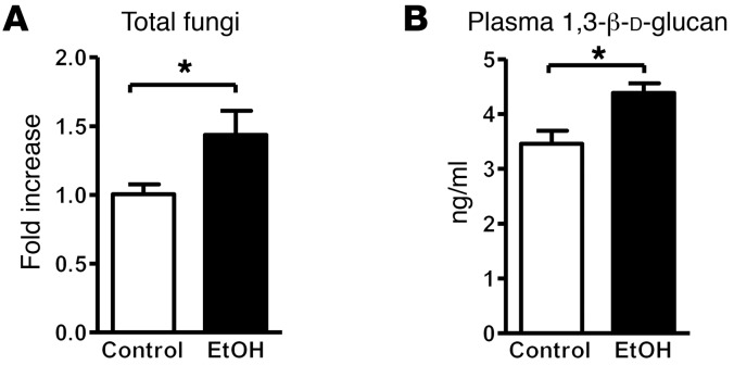
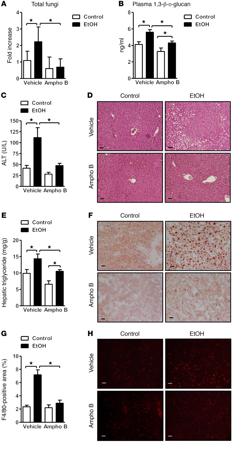
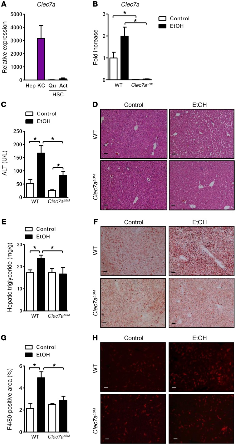
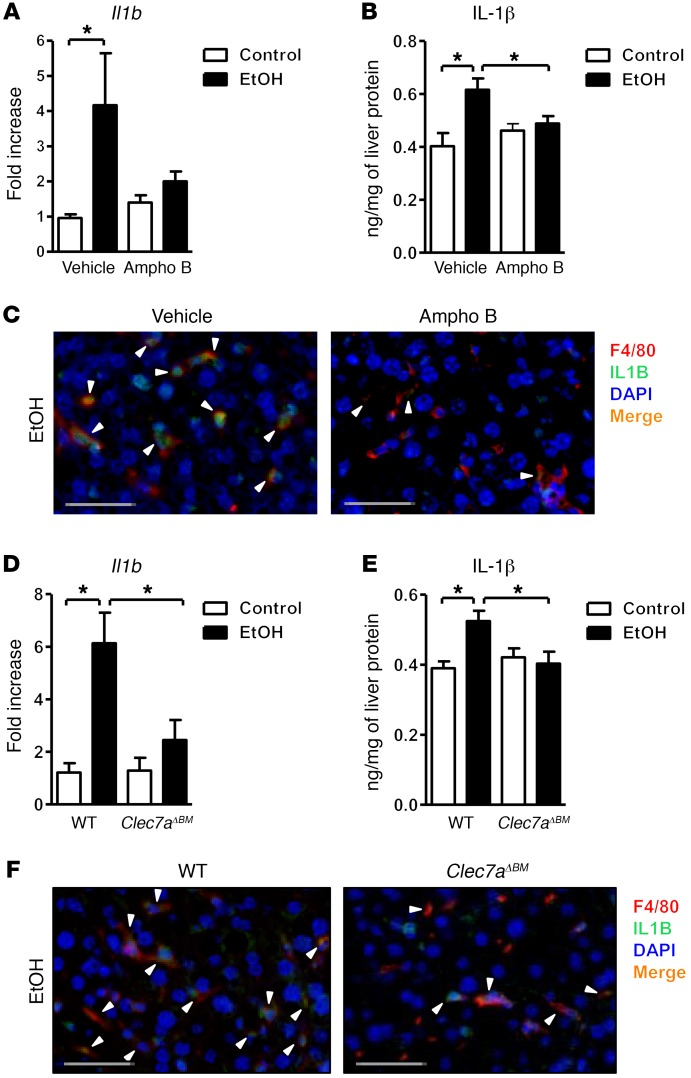
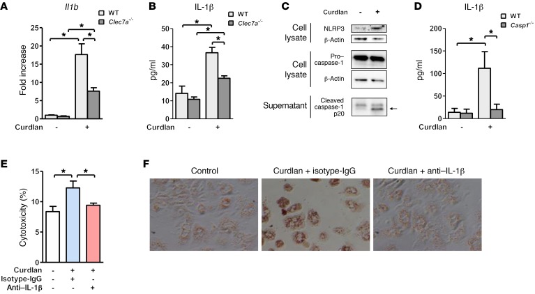
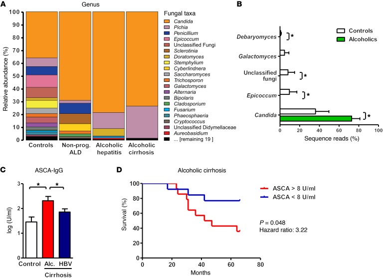
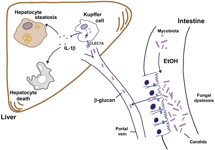
Chronic liver disease with cirrhosis is the 12th leading cause of death in the United States, and alcoholic liver disease accounts for approximately half of all cirrhosis deaths. Chronic alcohol consumption is associated with intestinal bacterial dysbiosis, yet we understand little about the contribution of intestinal fungi, or mycobiota, to alcoholic liver disease. Here we have demonstrated that chronic alcohol administration increases mycobiota populations and translocation of fungal β-glucan into systemic circulation in mice. Treating mice with antifungal agents reduced intestinal fungal overgrowth, decreased β-glucan translocation, and ameliorated ethanol-induced liver disease. Using bone marrow chimeric mice, we found that β-glucan induces liver inflammation via the C-type lectin-like receptor CLEC7A on Kupffer cells and possibly other bone marrow-derived cells. Subsequent increases in IL-1β expression and secretion contributed to hepatocyte damage and promoted development of ethanol-induced liver disease. We observed that alcohol-dependent patients displayed reduced intestinal fungal diversity and Candida overgrowth. Compared with healthy individuals and patients with non-alcohol-related cirrhosis, alcoholic cirrhosis patients had increased systemic exposure and immune response to mycobiota. Moreover, the levels of extraintestinal exposure and immune response correlated with mortality. Thus, chronic alcohol consumption is associated with an altered mycobiota and translocation of fungal products. Manipulating the intestinal mycobiome might be an effective strategy for attenuating alcohol-related liver disease.


Conflict of interest statement

Conflict of interest: The authors have declared that no conflict of interest exists.

Figures

Figure 1
Figure 1. Chronic alcohol feeding increases the number of fungi in the intestine and translocation of fungal products.
C57BL/6 mice were fed an oral control diet (n = 6–8) or ethanol diet (n = 8–14). (A) Total fungi in feces were assessed by qPCR. (B) Mean plasma levels of 1,3-β-d-glucan. Unpaired Student’s t test. *P < 0.05.
Figure 2
Figure 2. Decreased ethanol-induced liver disease in mice treated with antifungals.
C57BL/6 mice were fed an oral control diet (n = 4–14) or ethanol diet (n = 10–23), and also given vehicle or amphotericin B (Ampho B). (A) Total fungi in feces were assessed by qPCR. (B) Plasma levels of 1,3-β-d-glucan. (C) Plasma levels of alanine aminotransferase (ALT). (D) Representative liver sections after H&E staining. (E) Hepatic triglyceride content. (F) Representative Oil Red O–stained liver sections. (G and H) Representative liver sections of F4/80 immunofluorescence staining; positively stained area was quantified by image analysis software (n = 2–5). Scale bars: 50 μm. Unpaired Student’s t test. *P < 0.05.
Figure 3
Figure 3. Mice lacking CLEC7A in bone marrow–derived cells are protected from ethanol-induced liver disease.
(A) Expression of Clec7a in primary mouse hepatocytes (Hep), Kupffer cells (KC), and quiescent and activated hepatic stellate cells (QuHSC and ActHSC) was measured by qPCR (n = 2–3 independent experiments). (BH) C57BL/6 mice underwent transplantation of WT or Clec7a–/– bone marrow (Clec7aΔBM) and were fed an oral control diet (n = 5) or ethanol diet (n = 8–10; 3 technical replicates). (B) Hepatic Clec7a mRNA expression. (C) Plasma levels of ALT. (D) Representative liver sections after H&E staining. (E) Hepatic triglyceride content. (F) Representative Oil Red O–stained liver sections. (G and H) Representative liver sections of F4/80 immunofluorescence staining; positively stained area was quantified by image analysis software (n = 5–7). Scale bars: 50 μm. Unpaired Student’s t test. *P < 0.05.
Figure 4
Figure 4. Fungal products induce IL-1β in Kupffer cells.
(AC) C57BL/6 mice were fed an oral control diet (n = 5–11) or ethanol diet (n = 13–15) and given vehicle or amphotericin B (Ampho B). (A) Hepatic expression of Il1b mRNA. (B) Hepatic levels of IL-1β protein. (C) Immunofluorescence analysis of F4/80 (red) and IL-1β (green) (representative liver sections); nuclei are blue. (DF) C57BL/6 mice underwent transplantation of WT or Clec7a–/– bone marrow (Clec7aΔBM) and were fed an oral control diet (n = 5) or ethanol diet (n = 8–10). (D) Hepatic expression of Il1b mRNA. (E) Hepatic levels of IL-1β protein. (F) Immunofluorescence analysis of F4/80 (red) and IL-1β (green) (representative liver sections); nuclei are blue. Arrowheads indicate double-positive cells. Scale bars: 50 μm. Unpaired Student’s t test. *P < 0.05.
Figure 5
Figure 5. Curdlan induction of IL-1β by Kupffer cells contributes to hepatocyte injury and steatosis.
(A and B) Primary mouse WT and Clec7a–/– Kupffer cells were stimulated with curdlan; graphs show expression of Il1b mRNA (A) and secretion of IL-1β (B) (n = 3–6 independent experiments). (C) Levels of cellular NLRP3 and caspase-1 and cleaved caspase-1 in supernatants of Kupffer cells with and without curdlan stimulation. (D) IL-1β secretion by WT or caspase-1–deficient Kupffer cells following curdlan stimulation (n = 4–8 independent experiments). (E and F) Conditioned medium from Kupffer cells (stimulated or not stimulated with curdlan) was transferred to primary mouse hepatocytes in the presence of control (IgG) or IL-1β neutralizing antibody. Hepatocyte cytotoxicity (E) and lipid accumulation, determined by Oil Red O staining (F) (n = 3 independent experiments performed in triplicate). Unpaired Student’s t test (A and B); Mann-Whitney U-statistic test (D); 1-way ANOVA with Newman-Keuls post-test (E). *P < 0.05.
Figure 6
Figure 6. Fungal dysbiosis and immune response in alcohol-dependent patients.
(A and B) ITS sequencing of fecal samples from controls (n = 8), alcohol-dependent patients with nonprogressive alcoholic liver disease (Nonprog. ALD; n = 10), alcoholic hepatitis (n = 6), or alcoholic cirrhosis (n = 4). (A) The graph demonstrates the average relative abundance of sequence reads in each genus for controls and for each of the 3 liver disease stages. (B) Green bars indicate the average relative abundance of sequence reads in a combined alcohol-dependent group (independent of liver disease stage, n = 20) that were significantly different from the controls (white bars, n = 8). Kruskal-Wallis rank-sum statistical test with YAP. *P < 0.05. (C) Serum level of ASCA in healthy individuals (controls, n = 14) or patients with alcohol-related cirrhosis (Alc. cirrhosis; n = 28) or chronic HBV-related cirrhosis (n = 43). One-way ANOVA with Newman-Keuls post-test. *P < 0.05. (D) Kaplan-Meier curve of liver-related mortality for patients with alcoholic cirrhosis. Patients were grouped according to their serum levels of ASCA. Fourteen patients had serum concentrations of ASCA greater than 8 U/ml (the median value), and 13 had concentrations below this value. One patient was lost during the follow-up period.
Figure 7
Figure 7. Contribution of the intestinal mycobiota to alcoholic liver disease.
Chronic consumption of ethanol increases the fungal population and causes dysbiosis of the mycobiota in the intestine (right). The fungal dysbiosis results in higher amounts of β-glucan translocating across a damaged gut barrier to the liver (left). Increased β-glucan binds to CLEC7A on hepatic Kupffer cells and induces the expression and secretion of IL-1β. This cytokine contributes to ethanol-induced liver inflammation, hepatocyte injury, and steatosis.

Comment in

References

    1. Lozano R, et al. Global and regional mortality from 235 causes of death for 20 age groups in 1990 and 2010: a systematic analysis for the Global Burden of Disease Study 2010. Lancet. 2012;380(9859):2095–2128. doi: 10.1016/S0140-6736(12)61728-0. - DOI - PMC - PubMed
    1. WHO. Global Status Report On Alcohol And Health 2014. World Health Organization website. http://www.who.int/substance_abuse/publications/global_alcohol_report/en/ Accessed April 10, 2017.
    1. Bode JC, Bode C, Heidelbach R, Dürr HK, Martini GA. Jejunal microflora in patients with chronic alcohol abuse. Hepatogastroenterology. 1984;31(1):30–34. - PubMed
    1. Yan AW, et al. Enteric dysbiosis associated with a mouse model of alcoholic liver disease. Hepatology. 2011;53(1):96–105. doi: 10.1002/hep.24018. - DOI - PMC - PubMed
    1. Hartmann P, et al. Deficiency of intestinal mucin-2 ameliorates experimental alcoholic liver disease in mice. Hepatology. 2013;58(1):108–119. doi: 10.1002/hep.26321. - DOI - PMC - PubMed