. 2017 Jul;49(7):1025-1034.
doi: 10.1038/ng.3871. Epub 2017 May 22.

Mutations in DZIP1L, which encodes a ciliary-transition-zone protein, cause autosomal recessive polycystic kidney disease

Affiliations

Mutations in DZIP1L, which encodes a ciliary-transition-zone protein, cause autosomal recessive polycystic kidney disease

Hao Lu et al. Nat Genet. 2017 Jul.

Abstract

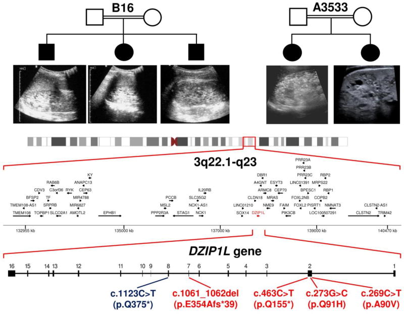
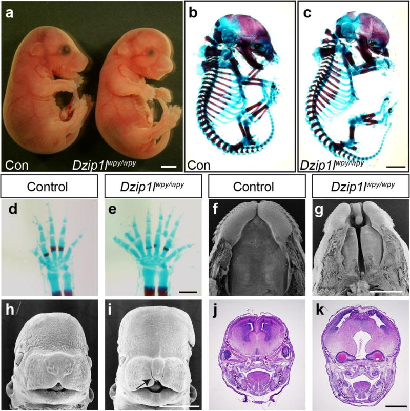
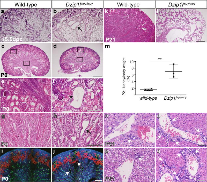
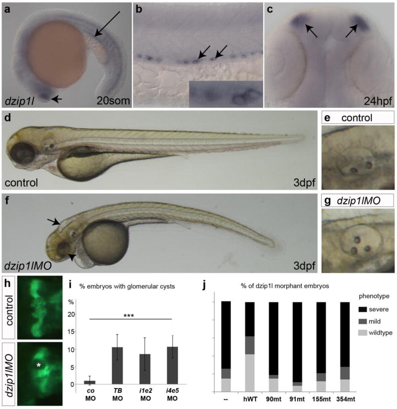
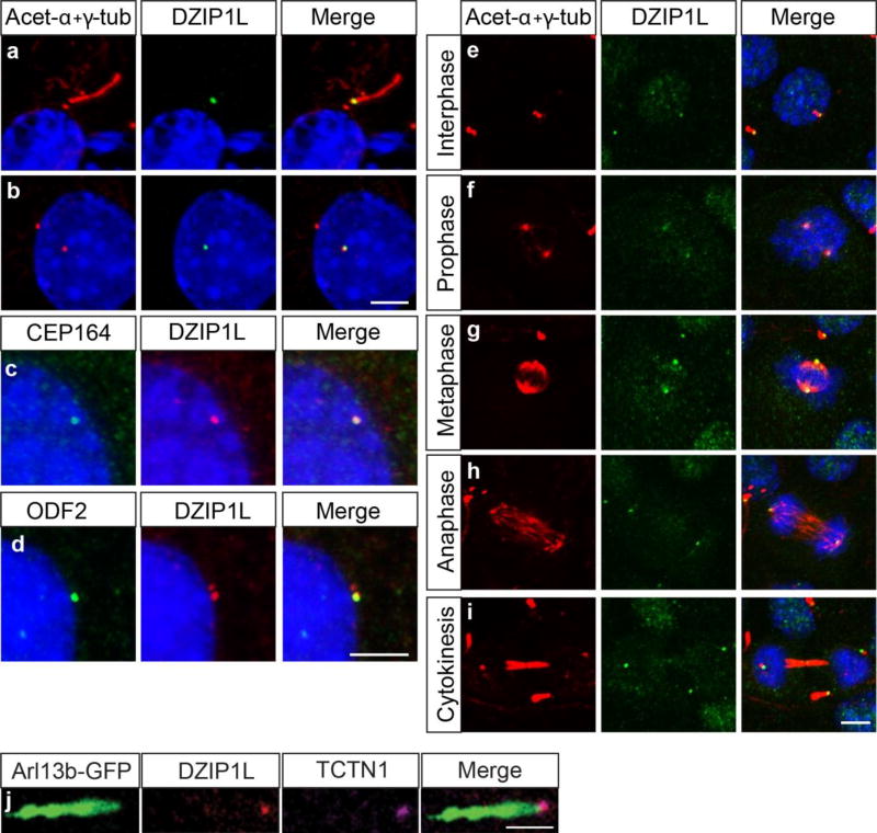
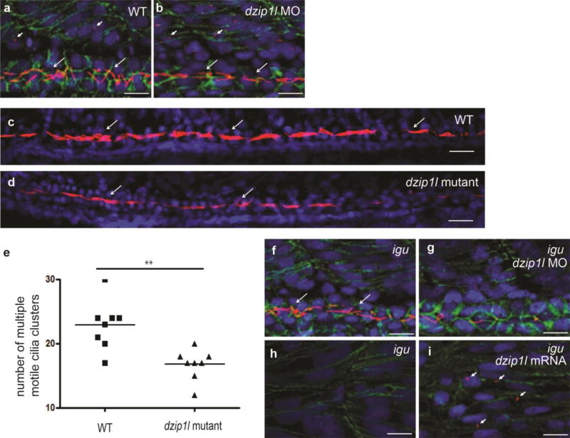
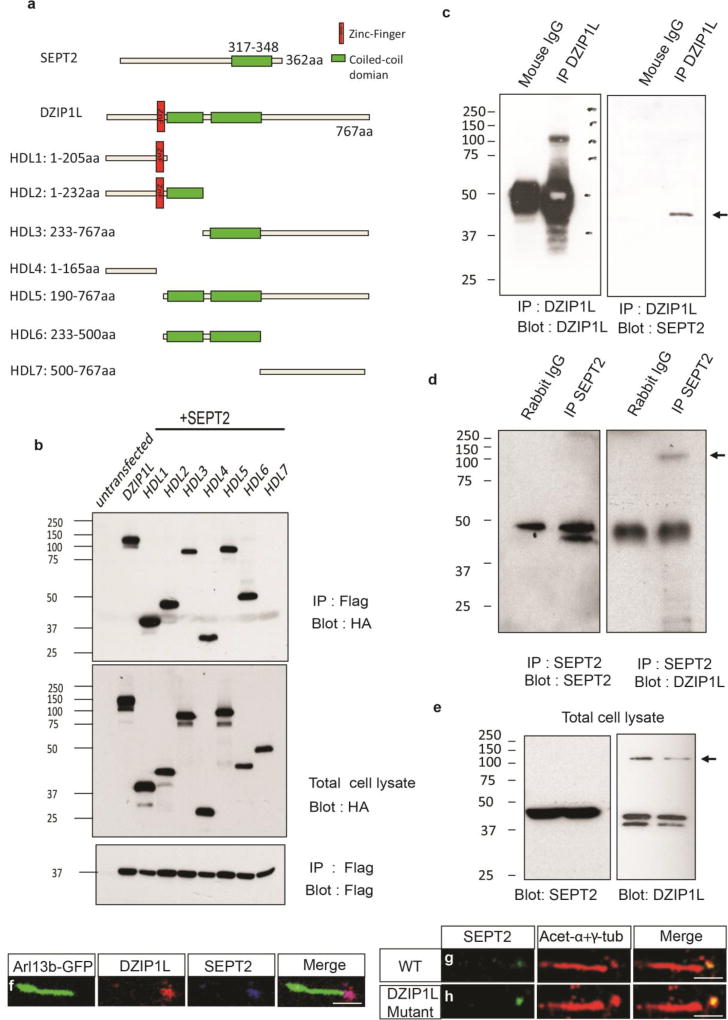
Autosomal recessive polycystic kidney disease (ARPKD), usually considered to be a genetically homogeneous disease caused by mutations in PKHD1, has been associated with ciliary dysfunction. Here, we describe mutations in DZIP1L, which encodes DAZ interacting protein 1-like, in patients with ARPKD. We further validated these findings through loss-of-function studies in mice and zebrafish. DZIP1L localizes to centrioles and to the distal ends of basal bodies, and interacts with septin2, a protein implicated in maintenance of the periciliary diffusion barrier at the ciliary transition zone. In agreement with a defect in the diffusion barrier, we found that the ciliary-membrane translocation of the PKD proteins polycystin-1 and polycystin-2 is compromised in DZIP1L-mutant cells. Together, these data provide what is, to our knowledge, the first conclusive evidence that ARPKD is not a homogeneous disorder and further establish DZIP1L as a second gene involved in ARPKD pathogenesis.


Figures

Figure 1
Figure 1
Mapping of a new ARPKD locus on 3q22.1-q23 and identification of DZIP1L mutations. Genome-wide SNP analysis performed in two unrelated consanguineous multiplex pedigrees (shown at top) with a total of five children affected by ARPKD, resulted in identification of a single overlapping 7.5 Mb region of homozygosity on chromosome 3q22.1-q23. By various sequencing approaches, we identified different homozygous DZIP1L mutations in these and other consanguineous families with ARPKD (see text for details). In parallel, we identified an ENU-induced recessive mouse model (see Fig. 2) bearing the homozygous Dzip1l nonsense mutation c.1123C>T (p.Gln375*) (in blue).
Figure 2
Figure 2
Dzip1lwpy/wpy mutant embryos show multiple dysmorphic features. (a) Control (left) and mutant (right) embryos at 17.5dpc. (b–e) Skeletal preps at 17.5dpc highlight defects in the craniofacial complex and polydactyly in Dzip1lwpy/wpy embryos (c,e) relative to controls (b,d). Dzip1lwpy/wpy mutant embryos consistently show some degree of cleft palate (f,g; arrowhead in g) and bilateral cleft lip (f-i; arrows in g,i). Scanning electron micrographs of 15.5dpc embryos shown with lower jaw removed in f,g; 13.5dpc embryo heads in h,i. (j,k) In a subset of embryos (n=30/45 scored) the eyes appeared abnormal, and in severe cases were buried within the skull (arrow in k). Images shown are of frontal sections through 15.5dpc embryonic heads. Note control embryos shown in (b,f,j) are heterozygous embryos that show no obvious phenotypic differences from wild-type controls, as shown in (a,d,h). The limbs shown in (d,e) are from different embryos than those shown in (b,c). Scale bars in a,b,c = 2mm; d,e = 0.5mm; f-k = 1mm. All data presented are from embryos on a C57BL/6:C3H mixed background.
Figure 3
Figure 3
Dzip1lwpy/wpy mice display progressive cystic kidney disease and hepatic defects following at least 4 crosses to a CD1 background. (a,b) Following H&E staining, cortical cysts were evident from 15.5dpc in Dzip1lwpy/wpy kidneys (arrow in b; n=4 independent embryos). (c,d,g,h) At P0, both cortical cysts (arrowhead in d,g) and medullary dilatations (arrow in d,h) were observed (n=6). (g,h) Show high power images of the regions marked in (d). (e,f,i,j) Immunofluorescence analysis of sections from P0 kidneys stained with Aqp2 (collecting ducts; green), and LTL-biotin (proximal tubules; magenta). (i,j) Show high power images of the proximal tubule cyst (i) and collecting duct dilatation (j) marked with an arrowhead and arrow respectively in (f). (k,l) By P21, cysts extend throughout the kidney, and (m) when corrected for body weight Dzip1lwpy/wpy kidneys are heavier than wild-type (n= 3 mutant, 4 wild-type embryos). Statistical analysis based on unpaired t-test with Welch’s correction to account for unequal variances, *p=0.05. Error bars show SEM. A significant difference was confirmed with a Student’s t-test on log transformed data, where the variances are equalized (not shown). (n–q) H&E staining of P21 liver sections. Arrows mark an excess of bile ducts, which nearly circumferentially surround portal vein branches in P21 Dzip1lwpy/wpy mutant (o,q) compared to wild-type (n,p) mice, indicating DPM (n=2). Scale bars in a,b = 100µm; c-f = 200µm, k,l = 500µm and g-j and n-q = 50µm.
Figure 4
Figure 4
DZIP1L localization overlaps with basal body, centrosome and transition zone markers. (a,b) IF staining for DZIP1L (green) and acetylated-α-tubulin and γ-tubulin (both in magenta) in serum starved IMCD3 kidney cells. DZIP1L staining overlaps with the basal body in ciliated cells (a) and the centrosome in non-ciliated cells (b). (c–d) Co-localization of DZIP1L (magenta) with the distal (CEP164, green in c) and subdistal (ODF2, green in d) appendage proteins. (e–i) DZIP1L (green) tracks with the centrioles (magenta) throughout all stages of the cell cycle in IMCD3 cells. (j) TCTN1 (magenta) and DZIP1L (cyan) co-localize at the transition zone in human dermal fibroblasts. ARL13B-GFP was used to mark the ciliary membrane around the axoneme (green in j). (k) 3D-SIM superresolution microscopy on RPE-1 (human retinal pigment epithelial) cells confirms both DZIP1L (magenta) and TCTN1 (cyan) are closely associated at the transition zone. Cilium labeled with ARL13B-GFP (green). Nuclei are stained with DAPI (blue). Scale bars in a-I = 5µm; j = 1µm; k = 500 nm. DZIP1L stained with Sigma C-terminal antibody, except in panels c,d,j,k where the Abnova antibody was used.
Figure 5
Figure 5
Dzip1l is required for ciliary differentiation in the zebrafish kidney tubule. (a) Primary cilia (short arrows; myotome) and mono motile cilia (long arrows; pronephric duct) in a 24 hpf wild-type embryo. (b) No obvious difference in primary (short arrows) and mono motile cilia (long arrows) differentiation in a dzip1l splice morpholino (e5i5) injected embryo at 24 hpf (n=10). (c) MCC motile cilia bundles (arrows) in the pronephric duct of a wild-type embryo at 48 hpf (arrows). (d) MCC motile cilia bundles (arrows) are reduced in a 48 hpf maternal-zygotic dzip1l mutant zebrafish embryo (n=8). (e) Quantification of MCC cilia bundles in wild-type and maternal-zygotic dzip1l mutant zebrafish embryos. 8 embryos from each group were analyzed. **p=0.0017 based on unpaired Student’s t-test. Error bars show SEM. In all panels, cilia were stained with anti-acetylated tubulin antibody (magenta) and nuclei with DAPI (blue); in panels a,b,f-i cell membranes were stained with β-catenin antibodies (green). Scale bars in a,b,f-i = 10µm; c,d = 100µm.
Figure 6
Figure 6
DZIP1L associates with the ciliary transition zone protein SEPT2. (a) Schematic diagram of SEPT2, DZIP1L and a series of DZIP1L deletion mutants. (b) Interaction between SEPT2 (N-terminal FLAG) and DZIP1L and various DZIP1L deletion mutants (C-terminal HA) as determined by IP following transfection in HEK293T cells. (c,d) Endogenous interaction between DZIP1L and SEPT2 in RPE-1 cells. IP was performed with either the anti-DZIP1L antibody (Abnova; c) or the anti-SEPT2 antibody (d). (e) Expression of DZIP1L and SEPT2 in total cell lysate. (f) Co-localization of DZIP1L (cyan) and SEPT2 (magenta) in human dermal fibroblasts. ARL13B-GFP was used to mark the ciliary axoneme (green). (g,h) SEPT2 (green) localization to the transition zone was unaffected in DZIP1L mutant dermal fibroblasts from individual B155 (p.Gln155*). Ciliary axonemes and basal bodies were labelled with anti-acetylated tubulin and anti-γ-tubulin antibodies (magenta), respectively. Scale bars in f-h = 1µm. All co-IP experiments were repeated at least three times.
Figure 7
Figure 7
Loss of DZIP1L affects the localization of PC1 and PC2 to the ciliary membrane. (a) In most wild-type ciliated MEFs PC1 (green; 7e12 mAb, Abcam) staining was distributed along the length of the axoneme as marked with ARL13B (magenta). (b) By contrast PC1 staining was more often concentrated at the proximal end of the axoneme in Dzip1lwpy/wpy MEFs (c) Quantification of the percentage of ciliated cells with PC1 along the axoneme (n=6, derived from 3 MEF cell lines from independent embryos, each counted in two separate experiments; between 50–185 cells counted per replicate, total 737 Dzip1l+/+ and 649 Dzip1lwpy/wpy cells counted). (d,e) Decreased PC1 (green) localization in the ciliary membrane in human dermal fibroblasts from affected individual B155 (p.Gln155*). ARL13B (magenta) marks the ciliary axoneme. (g,h) Decreased PC2 localization (green) in the ciliary membrane in DZIP1L mutant human dermal fibroblasts. Acetylated-α–tubulin and γ-tubulin (magenta) mark the cilia. (f,i) Quantification of the percentage of ciliated cells with PC1 and PC2 along the axoneme, respectively, in control and DZIP1L mutant human dermal fibroblasts. For human cells, cilia were counted in three experiments, with cells from three independent coverslips counted for each experiment (approximately 100 ciliated cells counted on each coverslip). In all cases, quantification included cilia with or without detectable PC1 or PC2 staining. Statistical analyses based on unpaired Student’s t-test. Error bars show SEM. For PC1 staining on MEFs, ****p<0.0001; PC1 and PC2 staining on human dermal fibroblasts, ***p<0.001. Scale bars in a,b = 2µm; d,e,g,h = 1µm.

References

    1. Guay-Woodford LM, et al. Consensus expert recommendations for the diagnosis and management of autosomal recessive polycystic kidney disease: report of an international conference. J Pediatr. 2014;165:611–7. - PMC - PubMed
    1. Bergmann C, et al. Clinical consequences of PKHD1 mutations in 164 patients with autosomal-recessive polycystic kidney disease (ARPKD) Kidney Int. 2005;67:829–48. - PubMed
    1. Habbig S, Liebau MC. Ciliopathies - from rare inherited cystic kidney diseases to basic cellular function. Mol Cell Pediatr. 2015;2:8. - PMC - PubMed
    1. Garcia-Gonzalo FR, et al. A transition zone complex regulates mammalian ciliogenesis and ciliary membrane composition. Nat Genet. 2011;43:776–84. - PMC - PubMed
    1. Jensen VL, et al. Formation of the transition zone by Mks5/Rpgrip1L establishes a ciliary zone of exclusion (CIZE) that compartmentalises ciliary signalling proteins and controls PIP2 ciliary abundance. EMBO J. 2015;34:2537–56. - PMC - PubMed

MeSH terms