Review
. 2017 Jul 31;46(15):4661-4708.
doi: 10.1039/c6cs00542j.

Self-assembling peptide and protein amyloids: from structure to tailored function in nanotechnology

Affiliations
Review

Self-assembling peptide and protein amyloids: from structure to tailored function in nanotechnology

Gang Wei et al. Chem Soc Rev. .

Abstract

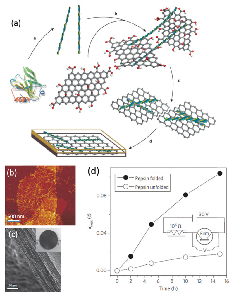
Self-assembled peptide and protein amyloid nanostructures have traditionally been considered only as pathological aggregates implicated in human neurodegenerative diseases. In more recent times, these nanostructures have found interesting applications as advanced materials in biomedicine, tissue engineering, renewable energy, environmental science, nanotechnology and material science, to name only a few fields. In all these applications, the final function depends on: (i) the specific mechanisms of protein aggregation, (ii) the hierarchical structure of the protein and peptide amyloids from the atomistic to mesoscopic length scales and (iii) the physical properties of the amyloids in the context of their surrounding environment (biological or artificial). In this review, we will discuss recent progress made in the field of functional and artificial amyloids and highlight connections between protein/peptide folding, unfolding and aggregation mechanisms, with the resulting amyloid structure and functionality. We also highlight current advances in the design and synthesis of amyloid-based biological and functional materials and identify new potential fields in which amyloid-based structures promise new breakthroughs.


Figures

Fig. 1
Fig. 1
Tapping mode AFM height images showing the morphology of C16-KTTKS at pH values of (a) pH 2, (b) pH 3, (c) pH 4, (d) pH 7. Reprinted with permission from ref. . Copyright 2013, Royal Society of Chemistry.
Fig. 2
Fig. 2
Schematic of the defibrillization of α-synuclein upon either cold or hot denaturing. Reprinted with permission from ref. . Copyright 2014, Wiley-VCH Verlag GmbH & Co.
Fig. 3
Fig. 3
Proposed modes of metal ion binding involved in the aggregation of the three proteins or peptides indicated. Reprinted with permission from ref. . Copyright 2012, Elsevier Ltd.
Fig. 4
Fig. 4
Formation of amyloid fibrils by α-synuclein. A solution was agitated in the presence of particles of hydrophobic PTFE, slightly hydrophilic PMMA or chemically inert glass; experiments with air injected at the top of the cuvette were also performed. Inset: Proposed mechanism of the fibrillization. The aggregation of proteins into fibrils (at the rate constant kfib), caused by association of the protein hydrophobic NAC domains, is enhanced in the presence of the hydrophobic PTFE interface. Reprinted with permission from ref. . Copyright 2010, Nature Publishing Group.
Fig. 5
Fig. 5
(a) Individual microscopic events underlying the aggregation mechanisms of amyloids. (b) The identification of the aggregation mechanisms and the specific intervention on targeted microscopic reactions are fundamental for the rational design of tailored functions. This concept is illustrated here with the example of the peptide Aβ1–42, for which the generation of particularly active species can be modulated by inhibiting different microscopic steps. Reprinted with permission from ref. . Copyright 2015 Nature Publishing Group.
Fig. 6
Fig. 6
Schematic assembly pathways of lysozyme oligomers at both denaturing and native temperatures. Reprinted with permission from ref. . Copyright 2014, American Chemical Society.
Fig. 7
Fig. 7
(a–c) Crystal structure of macrocyclic peptides with (a) monomer, (b) dimer, and (c) tetramer. (d) Several interaction modes of dimers to form a tetramer. Reprinted with permission from ref. . Copyright 2011 American Chemical Society.
Fig. 8
Fig. 8
Typical strategies for creating 0D amyloid (a) nanoparticles and (b) nanoclusters: (a) Aβ oligomer self-aggregation, (b) lipid bilayer membrane-induced Aβ assembly. CTB is cholera toxin B subunit. ASIGN is Aβ-sensitive ganglioside nanocluster. Images (a and b) are reproduced with permission from (a) ref. , Copyright 2014, American Chemical Society, and (b) ref. , Copyright 2013, American Chemical Society.
Fig. 9
Fig. 9
Amyloid triangles, squares and loops of ApoC-III. (a and b) TEM image of loops and the electron diffraction pattern, (c) AFM image of loops and (d) TEM images of triangles and polyhedra. Reproduced with permission from ref. . Copyright 2014, American Chemical Society.
Fig. 10
Fig. 10
Individual protofilaments of β-lactoglobulin aligning and starting to attach at specific points to form first protofibrils and finally mature fibrils. Reproduced with permission from ref. . Copyright 2011 Royal Society of Chemistry.
Fig. 11
Fig. 11
Twisted and helical amyloid ribbons: (a) left-handed β-lactoglobulin nanofibrils with multistranded twisted filaments. Reprinted with permission from ref. . Copyright 2010, Nature Publisher. (b) Cross-β amyloid TTR105–115 fibril with triplet atomic-resolution structure. Reprinted with permission from ref. . Copyright 2013, National Academy of Sciences. (c) Twisted right-handed helical ILQINS hexapeptide ribbon. Reproduced with permission from ref. . Copyright 2014, American Chemical Society. (d) Twisted double-helical peptide ribbon, Reprinted with permission from ref. . Copyright 2009, Wiley-VCH Verlag GmbH & Co. (e) Amyloid-inspired rippled β-sheet ribbons by the co-assembly of enantiomeric amphipathic peptides. Reprinted with permission from ref. . Copyright 2012, American Chemical Society.
Fig. 12
Fig. 12
Multistranded amyloid ribbons: (a) Tau protein R3 ribbon, reprinted with permission from ref. . Copyright 2016 Wiley-VCH Verlag GmbH & Co. (b) Multistranded hIAPP20–29 ribbon, reprinted with permission from ref. . Copyright 2013, National Academy of Sciences. (c and d) Lysozyme (c) and β-lactoglobulin (d) ribbons, reprinted with permission from ref. . Copyright 2011, American Chemical Society. (e) Amelogenin ribbon, reprinted with permission from ref. . Copyright 2016, Nature Publishing Group.
Fig. 13
Fig. 13
(a–c) Supramolecular co-assembly for the formation of amyloid peptide nanotubes with controllable dimensions: (a) co-assembly mechanism of FF and Boc-FF, (b) SEM image of peptide nanotubes with a FF/Boc-FFF ratio of 5 : 1 and (c) length distribution. Reproduced with permission from ref. . Copyright 2016, American Chemical Society. (d and e) Co-assembly of amyloid amphiphilic peptide-based molecules to form multiwalled nanotubes: (d) co-assembly mechanism, and (e) TEM image of peptide nanotubes. Reproduced with permission from ref. . Copyright 2014, American Chemical Society.
Fig. 14
Fig. 14
Typical methods for the fabrication of 2D protein amyloids: (a) stacking, (b) filtration and (c) self-assembly at an air–water interface. Image (a–c) are reproduced by permission from (a) ref. , Copyright 2010, Nature publishing Group, (b) ref. , Copyright 2015, American Chemical Society, and (c) ref. , Copyright 2016, Royal Society of Chemistry.
Fig. 15
Fig. 15
(a) Schematic illustration of the proposed mechanism for amyloid nanofilm formation. (b–d) Schematic strategies for lysozyme fibril nanofilm formation: (b) on the surface of immersed materials (solid–liquid interface) and (c) at the aqueous solution surface (vapour–liquid interface). (d) The contact-printing technique to deposit the free-floating amyloid nanofilm onto water-sensitive substrates. Reproduced with permission from ref. . Copyright 2015, Wiley-VCH Verlag GmbH & Co.
Fig. 16
Fig. 16
(a) Amino acid sequence of Aβ1–42 and the molecular structure of Aβ16–22; (b) AFM image and height analysis of self-assembled amyloid nanosheets; (c) structural model of the KLVFFAK nanosheet. Reproduced with permission from ref. . Copyright 2015, National Academy of Sciences.
Fig. 17
Fig. 17
Synthesis of amyloid 3D lysozyme microgels from amyloid fibril networks: (a) water-in-oil microgels, and (b) oil-in-water microgels. (c–f) Typical (c and d) 3D reconstructions of the confocal images and (e and f) cryo-SEM images of: (a) water-in-oil and (b) oil-in-water microgels, respectively. Reproduced with permission from ref. . Copyright 2015, American Chemical Society.
Fig. 18
Fig. 18
Length scale of various amyloid structures from monomer to oligomers, 0D, 1D, 2D and 3D.
Fig. 19
Fig. 19
(a) Formation mechanism of curli fibrils in E. coli biofilm. Reprinted with permission from ref. . Copyright 2008, Wiley-VCH. (b) 3D projection (upper) and side view (lower) CLSM images of S. typhimurium biofilms with a growth period of 24, 48 and 72 h. (c) CLSM image of a 72 h biofilm with DNA staining. Reproduced with permission from ref. . Copyright 2015, Elsevier Inc.
Fig. 20
Fig. 20
(a–c) Amyloid protein nanofibre hydrogel: (a) formation mechanism, (b) SEM and (c) fluorescence images. Reproduced with permission from ref. . Copyright 2010 Elsevier Ltd. (d–f) Amyloid peptide nanofibre hydrogel: (d) formation mechanism, (e) optical image of hydrogel and (f) AFM image. Reproduced with permission from ref. . Copyright 2015 Elsevier Ltd.
Fig. 21
Fig. 21
(a) Atomistic model of a PEG chain docked to Aβ1–42. The chain interacts with both hydrophobic as well as hydrophilic residues of the peptide and forms a spiral structure. (b) Best 50 conformations of the PEG chain (purple) docked to Aβ1–42 (orange), and (c) alkyl PE chain (purple) docking on Aβ1–42 (orange). Images (a–c) are reproduced with permission from ref. . Copyright 2012, American Chemical Society. (d–g) AFM (bottom row) images of the β-lactoglobulin amyloid fibrils (d) and fibrils decorated by gold (e), silver (f) and palladium (g) nanoparticles after the respective metal salt reduction by NaBH4. Images (d)–(g) are reproduced with permission from ref. . Copyright 2014, American Chemical Society.
Fig. 22
Fig. 22
(a) Schematic representation of the GQDs used for inhibiting the aggregation of Aβ1–42 peptides. (b) The kinetics of Aβ1–42 aggregation as monitored by the thioflavin T fluorescence in the absence or presence of GQD. Reproduced with permission from ref. . Copyright 2015 Royal Society of Chemistry. (c) AFM images of the fibrils obtained from mixtures of (i–iii) QD–bAS (QD/bAS = 1/40), and (iv) α-synuclein alone. Scale bars: 10 μm. Reproduced with permission from ref. . Copyright 2009, American Chemical Society.
Fig. 23
Fig. 23
(a) Schematic representation of the fabrication of bone-mimetic composites based on amyloid fibril and HA platelets. (b) TEM image of β-lactoglobulin amyloid fibrils. (c–e) SEM image of HA platelets (c), surface (d) and fracture sections (e) of the amyloid-based composite with 60 wt% brushite platelets. Reproduced with permission from ref. . Copyright 2014, Wiley-VCH Verlag GmbH & Co.
Fig. 24
Fig. 24
Formation of amyloid nanofibril–MWNT hybrid: (a) schematic illustrations of the functionalization of CNTs with amyloid fibrils. (b–d) TEM images of hybrids consisting of functionalized MWNTs (black arrows) and amyloid fibrils (white arrows) at 0.1 wt% with: (b and c) covalent and (d) non-covalent functionalization. Reproduced with permission from ref. . Copyright 2012, American Chemical Society.
Fig. 25
Fig. 25
(a) CR and NBT assays confirm the amyloidogenic features and the formation of an amyloid–DOPA hybrid. (b) TEM images of unmodified and modified amyloid nanofibrils with biomolecules. (c) Schematic presentation of the measurement of the adhesion force between amyloid nanofibrils and biomolecules with AFM force spectroscopy. Representative AFM image showing modified Mfp5-CsgA fibres 1 h after deposition on a mica surface. Reproduced with permission from ref. . Copyright 2014, Nature Publishing Group.
Fig. 26
Fig. 26
(a) Schematic orientation of β-sheets and β-strands in silk fibroin fibrils and amyloid fibrils. (b) Schematic illustration and cross-polarized light observation image of silk fibroin fibrils and amyloid fibrils composite film. (c–e) 2D-WAXS patterns of the films containing: (c) 100% silk fibroin fibrils, (d) 100% amyloid fibrils and (e) 5 : 5 silk fibroin fibrils : amyloid fibrils. (f) Magnetic functionalization and tensile properties of the film (silk fibroin fibrils : amyloid fibrils : magnetic nanoparticles weight ratio of 70 : 10 : 20), as prepared by vacuum filtration. (g) Shape-memory properties of the magnetic composite film when exposed to the combined presence of an external magnetic field and water. Reproduced with permission from ref. . Copyright 2014 Wiley-VCH Verlag GmbH & Co.
Fig. 27
Fig. 27
(a) Layered organization of amyloid fibrils and gold platelet hybrid nanocomposites. Reproduced with permission from ref. . Copyright 2013, Wiley-VCH Verlag GmbH & Co. (b) An illustrative description of the development of a photoluminescent peptide–QD hydrogel through the self-assembly of Fmoc-FF building blocks and their PL quenching associated with the enzymatic detection of analytes. Reprinted with permission from ref. . Copyright 2014, Wiley-VCH Verlag GmbH & Co.
Fig. 28
Fig. 28
(a) Schematic representation of the internalization and transport of metal nanoparticle-decorated amyloid fibrils into living cancer cells. Reprinted with permission from ref. . Copyright 2014, American Chemical Society. (b) Proposed coating mechanism of SEVI fibrils with amyloid-binding oligomers. These coatings prevent the direct interaction of HIV-1 with SEVI fibrils and prevent SEVI-mediated enhancement of viral infection in cells. Reprinted with permission from ref. . Copyright 2012, American Chemical Society.
Fig. 29
Fig. 29
Amyloid hydrogels for cell culture: (a) schematic of amyloid hydrogels for 2D and 3D cell cultures. (b) Schematic of the morphology of cells at each stage during the implantation. Stage 1: cultured cells for 24 h; stage 2: cells were primed with a differentiation medium for 5 days; stage 3: cells were then transplanted with hydrogel A5 into the mice brains, and stage 4: the brains were harvested. Scale bars: 200 μm for stage 1–3 and 100 μm for stage 4. (c) Implanted GFP-hMSCs with α-synuclein hydrogel (left) and without a hydrogel (right) at the caudate putamen after 7 days in vivo. (d) Cell viability when implanted with and without a hydrogel. (e) Box plot of the area with survived cells when transplanted with and without a hydrogel. Reproduced with permission from ref. . Copyright 2016, Nature Publishing Group.
Fig. 30
Fig. 30
(a) Biomimetic photosynthesis by light-harvesting peptide nanotubes. Reproduced with permission from ref. . Copyright 2012, Wiley-VCH Verlag GmbH & Co. (b) Schematic diagram of the hybrid photovoltaic device prepared using an active layer composed of TiO2-hybrid nanowires blended with polythiophene and AFM image of TiO2 decorating the surface of the amyloid fibrils. Reproduced with permission from ref. . Copyright 2012, Wiley-VCH Verlag GmbH & Co.
Fig. 31
Fig. 31
Amyloid nanofibril-based materials for water purification. (a) Structure of the β-lactoglobulin protein with the heavy metal-binding motif highlighted, 121-cys, with a lead ion attached and the 121-cys-containing fragment (LACQCL) from β-lactoglobulin with docked Pb2+. (b and c) Concentrations of heavy metal and radioactive pollutants before and after filtration through the amyloid fibril-activated carbon hybrid adsorber membrane: (b) potassium dicyanoaurate(i); (c) mercury chloride. Reproduced with permission from ref. . Copyright 2016, Nature Publishing Group.
Fig. 32
Fig. 32
Fabrication of amyloid-based transistors. (a and b) PEDOT-S amyloid nanofibrils-based transistor: (a) molecular structure and (b) schematic picture of an electrolyte gated transistor. Reproduced with permission from ref. . 2008, American Chemical Society. (c and d) Amyloid–PAni hybrid nanofibril-based transistor: (c) synthesis and AFM image of PNF–PAni hybrid fibrils and (d) deposited hybrid fibrils on gold electrode array for conductivity measurements (PNF: peptide nanofibrils). Reproduced with permission from ref. . Copyright 2015, American Chemical Society.
Fig. 33
Fig. 33
Amyloid protein nanofibril-based immunosensors. (a) Schematic synthesis of Sup35-BAP nanofibrils. (b) Fabrication of an immunosensor architecture via biotin–streptavidin interaction. (c) Improved sensing performance compared to non-fibril sensors. Reproduced with permission from ref. . Copyright 2010, Elsevier Ltd.
Fig. 34
Fig. 34
Amyloid nanofibril–GO shape-memory materials. (a) Fabrication mechanism, (b) AFM of nanofibril–GO hybrid, (c) SEM image of nanofibril–GO hybrid film. (d) Biosensors for enzymatic activity measurements. Reproduced with permission from ref. . Copyright 2012 Nature Publishing Group.

References

    1. Aguzzi A, O’Connor T. Nat Rev Drug Discovery. 2010;9:237–248. - PubMed
    1. Weber KT, Sun Y, Tyagi SC, Cleutjens JPM. J Mol Cell Cardiol. 1994;26:279–292. - PubMed
    1. Cen L, Liu W, Cui L, Zhang WJ, Cao YL. Pediatr Res. 2008;63:492–496. - PubMed
    1. Reymann AC, Boujemaa-Paterski R, Martiel JL, Guerin C, Cao WX, Chin HF, De La Cruz EM, Thery M, Blanchoin L. Science. 2012;336:1310–1314. - PMC - PubMed
    1. Knowles TPJ, Vendruscolo M, Dobson CM. Nat Rev Mol Cell Biol. 2014;15:496. - PubMed