

Blue-Shifted Green Fluorescent Protein Homologues Are Brighter than Enhanced Green Fluorescent Protein under Two-Photon Excitation
- PMID: 28530831
- PMCID: PMC5474692
- DOI: 10.1021/acs.jpclett.7b00960
Blue-Shifted Green Fluorescent Protein Homologues Are Brighter than Enhanced Green Fluorescent Protein under Two-Photon Excitation
Abstract
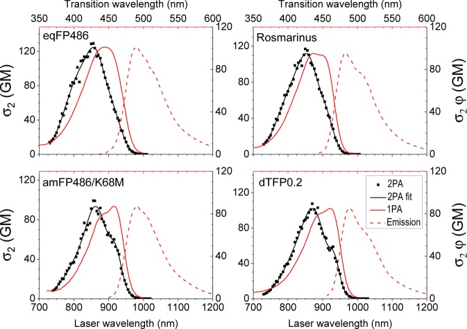
Fluorescent proteins (FPs) are indispensable markers for two-photon imaging of live tissue, especially in the brains of small model organisms. The quantity of physiologically relevant data collected, however, is limited by heat-induced damage of the tissue due to the high intensities of the excitation laser. We seek to minimize this damage by developing FPs with improved brightness. Among FPs with the same chromophore structure, the spectral properties can vary widely due to differences in the local protein environment. Using a physical model that describes the spectra of FPs containing the anionic green FP (GFP) chromophore, we predict that those that are blue-shifted in one-photon absorption will have stronger peak two-photon absorption cross sections. Following this prediction, we present 12 blue-shifted GFP homologues and demonstrate that they are up to 2.5 times brighter than the commonly used enhanced GFP (EGFP).
Conflict of interest statement
The authors declare no competing financial interest.
Figures


Similar articles
-
Comparative photophysical properties of some widely used fluorescent proteins under two-photon excitation conditions.Spectrochim Acta A Mol Biomol Spectrosc. 2021 Dec 5;262:120133. doi: 10.1016/j.saa.2021.120133. Epub 2021 Jun 30. Spectrochim Acta A Mol Biomol Spectrosc. 2021. PMID: 34243141
-
Describing two-photon absorptivity of fluorescent proteins with a new vibronic coupling mechanism.J Phys Chem B. 2012 Feb 9;116(5):1736-44. doi: 10.1021/jp211020k. Epub 2012 Jan 30. J Phys Chem B. 2012. PMID: 22224830 Free PMC article.
-
Novel uses of fluorescent proteins.Curr Opin Chem Biol. 2015 Aug;27:1-9. doi: 10.1016/j.cbpa.2015.05.002. Epub 2015 May 25. Curr Opin Chem Biol. 2015. PMID: 26022943 Review.
-
Hidden electronic excited state of enhanced green fluorescent protein.J Phys Chem B. 2008 Mar 13;112(10):2761-3. doi: 10.1021/jp711628u. Epub 2008 Feb 15. J Phys Chem B. 2008. PMID: 18275187
-
New red-fluorescent calcium indicators for optogenetics, photoactivation and multi-color imaging.Biochim Biophys Acta. 2014 Oct;1843(10):2284-306. doi: 10.1016/j.bbamcr.2014.03.010. Epub 2014 Mar 27. Biochim Biophys Acta. 2014. PMID: 24681159 Review.
Cited by
-
Liver-secreted fluorescent blood plasma markers enable chronic imaging of the microcirculation.Cell Rep Methods. 2022 Sep 21;2(10):100302. doi: 10.1016/j.crmeth.2022.100302. eCollection 2022 Oct 24. Cell Rep Methods. 2022. PMID: 36313804 Free PMC article.
-
Engineering rhodopsins' activation spectra using a FRET-based approach.Biophys J. 2022 May 3;121(9):1765-1776. doi: 10.1016/j.bpj.2022.03.024. Epub 2022 Mar 21. Biophys J. 2022. PMID: 35331688 Free PMC article.
-
Comparing the performance of mScarlet-I, mRuby3, and mCherry as FRET acceptors for mNeonGreen.PLoS One. 2020 Feb 5;15(2):e0219886. doi: 10.1371/journal.pone.0219886. eCollection 2020. PLoS One. 2020. PMID: 32023253 Free PMC article.
-
NaNuTrap: a technique for in vivo cell nucleus labelling using nanobodies.Development. 2021 Sep 15;148(18):dev199822. doi: 10.1242/dev.199822. Epub 2021 Sep 1. Development. 2021. PMID: 34328170 Free PMC article.
-
What is the Optimal Size of the Quantum Region in Embedding Calculations of Two-Photon Absorption Spectra of Fluorescent Proteins?J Chem Theory Comput. 2020 Oct 13;16(10):6439-6455. doi: 10.1021/acs.jctc.0c00602. Epub 2020 Sep 21. J Chem Theory Comput. 2020. PMID: 32862643 Free PMC article.
References
MeSH terms
Substances
Grants and funding
LinkOut - more resources
Full Text Sources
Other Literature Sources
Research Materials
Miscellaneous
